
MARLOES SJOERDSMA
PROJECT: MULTI-PERSPECTIVE FUNCTIONAL ECHOCARDIOGRAPHY
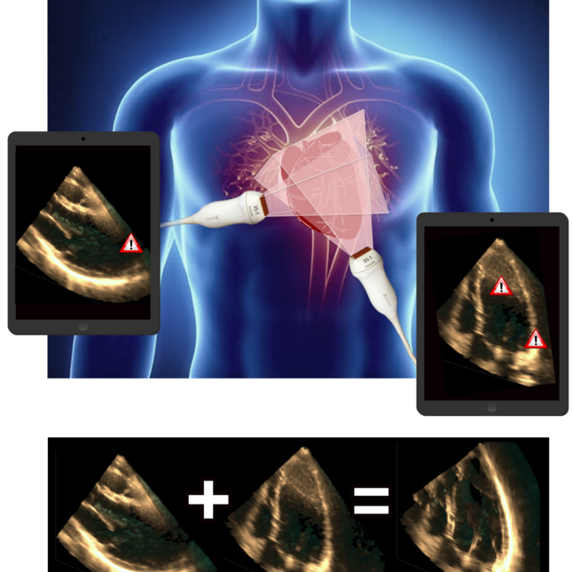
Current clinical guidelines for aortic valve stenosis diagnosis and intervention planning focus mainly on the pressure gradient over the valve, and on ejection fraction of the left ventricle. However, ejection fraction is merely applicable in case of global, late-stage cardiac disorders. Accordingly, left ventricular strain measured using ultrasound is a more suitable parameter. Although, time-resolved 3D ultrasound can capture the complex cardiac shape and contraction pattern, 3D+t ultrasound has a limited field-of-view, often contains near-field clutter, and has a relatively low lateral contrast and resolution.
Multi-perspective ultrasound (i.e. obtaining, registering, and fusing multiple ultrasound acquisitions) increases the field-of-view, and enhances the lateral contrast and resolution. In addition, a near-field clutter reduction algorithm was created and implemented in the fusion algorithm.
Thanks to the collaboration between the TU/e, Philips, and the cardiology and radiology department of the Catharina Hospital, high-quality ultrasound images could be recorded in the Catharina Hospital that were registered using a probe-tracker designed by Philips. After fusion of the images, 3D left ventricular strains could be obtained and compared to tagged-MRI (the golden standard for strain estimation). The collaboration was especially successful due to the face-to-face contact moments, during e.g. the weekly (pre-covid) visits to Philips Research and the meetings with healthcare professionals.
PEIRAN CHEN, ANDREAS POLLET, MEIYI ZHOU
PROJECT: INTEGRATED PLATFORM TO DESIGN NOVEL CANCER LOCALIZATION STRATEGIES BY ULTRASOUND MICROVASCULATURE IMAGING (LOCATE)
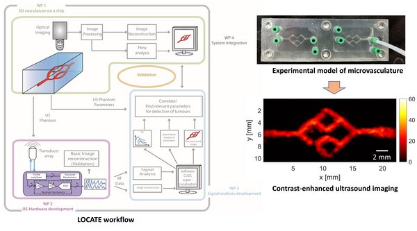
Each year, about 18 million new cancer cases and 9.5 million cancer-related deaths occur worldwide. Tumor-driven angiogenesis is a recognized hallmark of cancer growth, resulting in a complex and irregular microvascular architecture localized within the tumorous tissue. Tumor blood perfusion patterns become thus distinguishable from those in normal tissue. However, the relationship between the underlying microvascular architecture and the resulting hemodynamics is still not well understood, which hampers the development of a cost-effective and precise imaging solution for cancer localization. To close this gap, the LOCATE project came into being, aiming at opening new avenues towards a diagnostic solution for cancer localization by imaging tumor-related vasculature using contrast-enhanced ultrasound (CEUS). The specific goal of the project is to extend fundamental knowledge through the development and application of a novel integrated validation and development platform. The platform consists of experimental models of the microvasculature (work package 1, Andreas Pollet), tailored ultrasound imaging hardware based on custom-designed integrated chips (work package 2, Meiyi Zhou), and dedicated ultrasound image analysis strategies (work package 3, Peiran Chen). With this novel platform, the relationship between the microvascular architecture and the flow dynamics, along with the resulting CEUS images and their quantification can be investigated, which facilitates the development of diagnostic imaging solutions.
Thanks to the e/MTIC ecosystem, we have benefited greatly from the collaboration between Philips and TU/e.

LAURY QUAEDACKERS PROJECT: EXPLORING AND MONITORING THE NARCOLEPSY SYMPTOM SPECTRUM
Narcolepsy is a rare neurological sleep disorder with excessive daytime sleepiness and cataplexy as most pronounced symptoms. Both in the diagnostic process and in treatment, these symptoms are the primary focus. On the other hand, there are indications that the impact of narcolepsy is also determined by associated symptoms, such as social, psychiatric and cognitive problems. In the absence of disease-specific measures that map the experienced burden of symptoms, we have developed the narcolepsy monitor. The narcolepsy monitor is an app that aims to monitor a wide range of narcolepsy symptoms over a longer period of time, paying attention to the experienced burden of a symptom. The narcolepsy monitor makes it possible to collect a lot of data in a relatively non-invasive way about the presence of symptoms, the burden these symptoms cause and the dynamics between symptoms over time. An initial analysis of the data supports the hypothesis that in addition to the traditional symptoms, less obvious symptoms are also common and contribute significantly to the perceived burden. Prof. dr. Sebastiaan Overeem (Center for Sleep Medicine Kempenhaeghe and TU/e Biomedical Diagnostics Group) and prof. Panos Markopoulos (TU/e) played a crucial role in the project; the TU/e design department was involved in the initial design. Under the e/MTIC umbrella, students and clinicians joined forces to realize a pilot version of the app.
Lizeth Gonzalez Carabarin
Project: Prediction of life-threatening arrythmias based on low-power-and-low-memory Deep Neural Networks (project EuroTech).
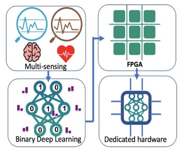
Among Machine Learning algorithms, Deep Learning (DL) is a powerful tool for classification and prediction. Unfortunately, current Deep Learning models remain costly in terms of power consumption and resources, significantly hampering its adoption in low-power wearables. Therefore, this work aims to design cutting-edge deep learning algorithms suitable for highly efficient hardware implementations. As a first demonstrator, the algorithm is used to predict life-threatening arrythmias based on ECG recordings. We leverage and optimize different model compression techniques (specifically pruning and quantization) based on a novel probabilistic approach to truly match the hardware needs. Based on our algorithms, DL architectures are compressed and are able to effectively classify ECG segments belonging to shockable and non-shockable arrhythmias. These ultra-compressed models are then suitable for hardware implementation and can be transferred to detect other life-threatening conditions.
This multidisciplinary project potentially opens different collaborations with industry and academia. On one side, this research benefits patients that requires real-time monitoring to detect life-threatening and dangerous events (e.g. life-threating arrythmias, epileptic seizure detection). On the other side, it requires the collaboration with hardware and ML experts (academia and/or industry) to co-optimize the DL models.
Ruben Deneer
Project: Extensive Personal Monitoring & Watch Platform (project ITEA eWatch)
The ITEA eWatch project is a transnational and industry driven R&D&I programme, with partners in Turkey, Belgium and the Netherlands. The goal is to develop an integrated platform focusing on the value of continuous physiological data in managing chronic diseases and monitoring patients’ post-hospitalisation recovery through wearable and/or medical devices. Coupled with the shift in healthcare towards prevention, the project targets the following technologies: pulse oximetry that establish real-time signal processing capabilities to filter out noise in order to continuously correct pulse oximetry data on a wearable, battery powered system; a wound/skin imaging system that allows nurses and patients to reliably image the wound or skin at the patient’s home and allow secure, reproducible real-time transmission of the images to a clinician in the hospital; indoor and outdoor localisation and security. In addition, the project will include data analysis solutions producing reports for doctors to be used in consultancy. Within the e/MTIC consortium there is a cooperation between Philips, the Eindhoven University of Technology (TU/e) and the Catharina Hospital. Philips provides wearable and sensing technologies, the TU/e intelligent systems development and the Catharina Hospital clinical expertise and access to its patient population through clinical trials.
Roel Meiburg
Project: Patient-specific pre-operative assessment of aortic stenosis using mathematical and physical models.
Patients suffering from calcific aortic stenosis experience an increased workload for the heart due to a significant narrowing of the aortic valve. The golden standard of treatment is valve replacement, but these patients are often elderly and suffer from one or more comorbidities – making them less suited to invasive procedures. Current diagnostic criteria focus mostly on the absolute valvular function (e.g., maximal pressure drop), mostly neglecting the cardiac and vascular function. Furthermore, the first symptoms often occur during exercise while diagnosis is performed at rest, assuming exercise physiology can be extrapolated from the resting state. Using silicone rubber valves with calcium deposits derived from patient measurements obtained at the Catharina Hospital Eindhoven, we showed that these assumptions do not hold. Instead, valve behaviour is highly heterogeneous and non-trivial. Applying this knowledge to computer models shows that this valvular heterogeneity significantly impacts the overall patient’s haemodynamics, and resting measures are not easily extrapolated to exercise. We formulated a new haemodynamic index (power transfer efficiency) which takes into account both cardiac and vascular function, which better describes the impact of the stenotic valve on the patient’s health. Finally, we used the silicone model to fully simulate valve replacement treatment, which could be used as a clinical training tool. By working directly with our clinical partners at the Catharina Hospital we were not only able to access unique, high-quality data, but also better understand the challenges in diagnosing these patients.
Tudor Văcărețu
Project: Smart subjective sleep monitoring in family context
One third of our lives is spent sleeping. Sleep is the activity that occupies most of our time. However, sleep is not a smooth experience for everyone. This can affect overall health, safety and quality of life. There is already a wide body of research around sleep, however, tracking sleep in a family context is still under researched.
This project investigates new ways and methods for tracking in situ family sleep. Both objective and subjective data are monitored through proposed software platforms and sensors which aim to capture patient data in an unobtrusive manner.
I am thankful to the e/MTIC ecosystem because I could approach this topic from different angles. At Kempenhaeghe I was allowed to observe meetings between patients and professionals, run interviews or focus groups.
Further, together with Philips Experience Design, during a one year study, sleep data was collected from five families to explore family sleep tracking at home. Last but not least, at TU/e, supported by Kempenhaeghe and Philips Experience Design I found the guiding I needed to carry out the research and a welcoming environment to work in.
Sophia E. Shanko
Project: Magnetic micromixing for point of care diagnostics
The coronavirus perfectly highlights the need of fast diagnostic tools that can be used at the point-of-care to enable fast decision making. Within these tools a drop of patient sample (i.e. blood) can be loaded where a target (like an antibody) can be found. A detection molecule (biosensor) is added that can detect the target, bind to it and change color. To ensure quick results, these tools can be further optimized. Instead of simply waiting for the biosensor to randomly encounter the antibody, their chances of meeting and binding can increase by introducing kinetics within the sample thereby mixing the contents of the chip. In our work we show that this enhancement can be realized by adding magnetic particles to the sample, which are moved in a controlled manner to induce mixing. We test our approach with a biosensor that detects HIV antibodies.
In specific biosensor-antibody conditions we can see a clear difference in time-to-result between mixing and non-mixing scenarios. Using such an approach in a point-of-care device we can halve the time of obtaining results clearly highlighting the benefits of mixing. In this way, the time needed to provide a diagnosis is decreased and fast decision making is enabled.
Yijing Zhang
Project: Enabling Widespread Ambulatory Monitoring for improved pregnancy outcome (EWAM)
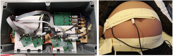
Non-contact capacitive electrodes can provide more comfort and have the biggest potential to be integrated into wearables, compared to conventional gel or dry electrodes, as they enable to measure biopotential signals through insulation layers (like clothes). This can be applied for fetal heart rate detection to build an unobtrusive and long-term monitoring method. However, dedicated hardware needs to be designed to handle the poor coupling quality and higher sensitivity to motion artefacts.
This project is part of the e/MTIC ecosystem. Thanks to the collaboration between MMC, Philips and TU/e, we designed and fabricated the front-end amplifier which is the most critical element in the signal chain. A 4-channel data acquisition system was built up with the IC and it has been tested with in-vivo experiments. Plausible results have been obtained with pregnant volunteers.
Ilde Lorato
Project: Camera-based Respiration Monitoring
Due to the respiratory instability in infants, continuous respiration monitoring is required in Neonatal Intensive Care Units (NICUs). Chest Impedance (CI) is commonly used in neonatal wards to monitor respiration. However, the electrodes attached to the infants’ skin can cause skin irritation and discomfort. Moreover, the method is sensitive to motion artifacts and unreliable for apnea detection.
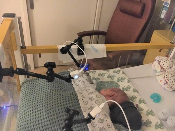
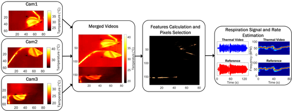
Camera-based respiration monitoring has been introduced as an unobtrusive alternative to CI, though problems with motion robustness and automatic detection of a region of interest remain. Thanks to the cooperation between MMC, Philips, and TU/e we built a setup and collected a dataset of thermal videos in the Medium Care Unit of MMC. We used three low-cost thermal cameras to record 15 infants for 3 hours. Thermal cameras were chosen because of the independence of light conditions and the visibility of both respiratory motion and flow. The videos are used to develop algorithms for respiration monitoring.
Our algorithm is based on an automatic pixels selection strategy. We developed three features that are able to identify the pixels containing the respiration information in both thermal and RGB/NIR videos. Our method does not require facial/body landmarks detection or skin visibility, broadening the possible application of this method in neonatal care.
This research is part of the ALARM project.
Bettine van Willigen
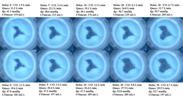
Project: Development of a digital twin of the artificial womb
To increase the survival chance of extremely preterm infants (born before 28 weeks of gestation), the Perinatal Life Support (PLS) project aims to develop an artificial womb (PLS system). This research develops a digital twin of the infant in the PLS system to aid clinicians in clinical decision-making.
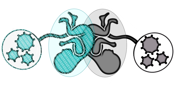
The digital twin of the artificial womb is a computer simulation of the fetus inside the womb (Fig.1). The purpose of this model is to gain more knowledge of the fetal physiology and to analyze the interaction of the components of the PLS system with each other and their effect on the fetus. Ultimately, this knowledge can be used as clinical decision support tool during PLS-based treatment by monitoring and interpreting vital signals of the fetus and inform the user to optimize (automatically) settings of the PLS system or support interventions by fetal distress.
When taking these purposes into account, the virtual fetus Arielle should contain physiological models of the cardiovascular system including autonomic regulation to simulate the hemodynamics of the fetus, the maternal-fetal gas exchange to simulate oxygen saturation, metabolism of the fetus, and fetal growth. In addition, the PLS components interacting with fetal physiology, such as the oxygenator, need to be modeled as well.
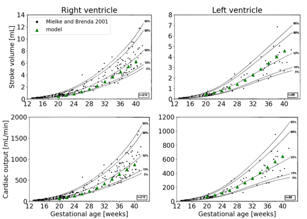
Working together with clinicians from Máxima Medical Center and Eindhoven University of Technology, the first version of Arielle is developed containing a fetal cardiovascular system and describes realistic cardiac output and stroke volume over gestational age from 20 to 40 weeks (Fig.2).
Mark van Gastel
Project: Camera-based vital signs monitoring during sleep
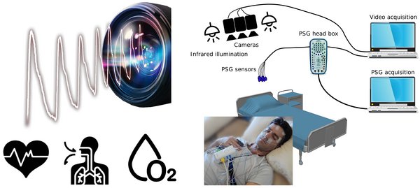
Sleep apnea is one of the most common sleep disorders affecting an estimated 9-21% of women and 24-31% of men worldwide. Polysomnography (PSG) is the current gold-standard tool for the diagnosis of sleep apnea. A collection of sensors is used to capture the physiological information required for a reliable diagnosis, which requires the patient to come to a specialized sleep hospital for a nocturnal observational study.
To improve the patient’s comfort and enable sleep diagnostics outside the clinic, we investigated if a camera combined with invisible near-infrared illumination can be used to monitor the key physiological parameters for the diagnosis of obstructive sleep apnea. The camera can detect the imperceptible skin color variations induced by blood volume variations, similar to photoplethysmography (PPG), but with a much smaller signal strength compared to a contact sensor, approximately 100 times. From the video data the physiological parameters pulse, respiration and peripheral oxygen saturation (SpO2) were automatically extracted of patients enrolled for PSG.
This project was part of the e/MTIC ecosystem, which enabled data collection with, and validation of the developed technology on patients at Kempenhaeghe. This research is continued, and further extended in the e/MTIC project UMOSA (Unobtrusive Monitoring of Obstructive Sleep Apnea).

Gabriele Varisco
Project: ALARM – Alarm-Limiting AlgoRithm-based Monitoring
Premature infants, born weeks too early, are treated in a neonatal intensive care unit (NICU), where their vital signs are continuously monitored. Alarms are used to indicate situations when a caregiver should attend to patients, but this can lead to a high alarm load. As a result caregivers can develop alarm fatigue, which can cause delayed responses to alarms, possibly resulting in patient harm.
By working in close contact with e/MTIC clinicians, engineers and industry, a new alarm management solution (PHASE-1) and subsequently an optimization program (PHASE-2) were introduced in the NICU of Máxima Medical Center to decrease the alarm burden. The latter included an alarm workshop, new training for nurses on alarm responses and revision of settings of oxygen saturation (SpO2) alarms.
Different measures were used to analyse alarms and vital parameters collected from October 2017 to December 2019, including the count of alarms and parallel alarms but also time spent by patients in different SpO2 ranges. Results showed a significant reduction in critical alarms and an increase in time spent by patients within the optimal SpO2 range (88%<SpO2<95%).
A survey was performed to evaluate how the system was perceived. The nurses responded that they believe that false alarms are less likely to cause an inappropriate response since the start of PHASE-2.
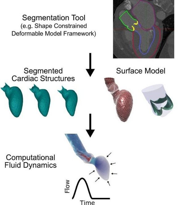
Martijn Hoeijmakers
Project: Analysis of aortic-valve blood flow using computational fluid dynamics
It is estimated that 2.5% of the population suffers from some form of heart valve disease. Studies suggest that aortic-valve stenosis – the narrowing of the aortic-valve opening area in systole – is most common and occurs in 43% of patients with valvular disease. The narrowing of the aortic-valve opening area imposes a large resistance to blood flow in systole. This is accompanied by a large (irreversible) transvalvular pressure-drop, which subsequently causes excessive loads on the heart. The transvalvular pressure-drop is an important clinical indicator of stenosis severity. However, accurate non-invasive estimation of this pressure-drop is challenging.
To resolve this, we combined medical imaging data with three-dimensional Computational Fluid Dynamics. Though such methods are time consuming and generally not feasible for clinical practice. This project used techniques such as statistical shape modeling and meta-modeling to make considerable steps to reduce simulation times and improve clinical feasibility.
This work was part of the EU-funded EurValve project which included various academic, clinical and industry partners among which e/MTIC partners such as Philips, the TU/e, and the Catharina Hospital. Even though the EurValve project has been completed for over two years, regular meetings with clinicians from the Catharina Hospital still provide valuable clinical feedback.
Laura Bogatu
Project: Faster and Better Detection of Deterioration of Post-Operative Patients using Non-Obtrusive Techniques
This project addresses the need for improved hemodynamic measurements in critical care. The main hypothesis is that the standard hemodynamic monitoring technology might not be used up to its full potential. The project explores how to adapt existing critical care technology for the purpose of developing new measurement strategies, aiming to obtain more complete information on the hemodynamic status of patients at risk of deterioration. We find that non-standard, but highly relevant parameters such as smooth muscle tone can be acquired via technologies already available in hospitals.
Our current approach aims at advanced understanding of cuff-based physiological measurements and characterization of circulatory system responses via occlusion-based perturbations. Methods include fusion of information acquired with established and common measurements such as non-invasive cuff-based blood pressure estimation, electrocardiogram, photo-plethysmogram, invasive blood pressure, but also imaging modalities such as MRI and ultrasound complemented by in silico simulations. Advanced understanding of the cuff measurement principle reveals insights for improved non-invasive blood pressure measurements and estimation of previously inaccessible hemodynamic parameters (e.g. smooth muscle tone, peripheral vascular properties). The work is conducted within the e/MTIC collaboration which facilitates such a multidisciplinary research approach involving data analytics, mechanical design, imaging, and technology development in proximity to the clinical environment.
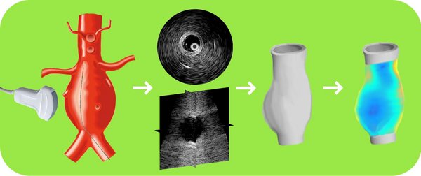
Floor Fasen
Project: Minimally Invasive Ultrasound Imaging of Abdominal Aortic Aneurysms
A localized widening in the largest artery in the human body, the aorta, can be life-threatening, when no preventive surgery is performed. This abdominal aortic aneurysm grows, and when the wall becomes too weak, rupture of the aortic wall may happen. Currently, the decision to do a surgical intervention is based on the aortic diameter and evidence-based medicine. However, the TU/e research group I am working in, already showed that there are better patient-specific markers to predict the risk of rupture based on mechanics.
The goal of this work is to utilize intravascular and 3D ultrasound to assess the thickness, geometry, and biomechanical state of the aneurysm by imaging from both the in- and outside of the aorta. Based on this new source of information, this work will focus on the development of new methods to predict the rupture risk, which could help to prevent unnecessary surgical risks. Currently, methods are being tested on porcine aortas. Soon, a clinical pilot study will be performed in the Catharina Hospital where I collaborate with vascular surgeons and sonographers. One day a week I work at Philips Research (outside of corona times), where I regularly speak to people working on similar technological challenges. This mix of academia, clinic and industry is very motivating.

RUBEN VAN DIJK
PROJECT: Nutrition monitoring with sensors in the home environment
Worldwide, millions of people suffer from cardiac diseases, leading to increasing pressure on healthcare systems. These cardiovascular diseases such as heart failure and atrial fibrillation are often the result of an unhealthy lifestyle. There is already much research into tracking physical activity and sleep, but tracking nutrition is relatively underexplored.
Previous work shows that letting people track what they eat and drink increases their awareness and can help them stick to a specific diet. However, the manual process of logging nutrition facts in a journal can be a tiring process for patients. As a result, people stop keeping track of their food, which lets them slip back into old habits.
This research explores new ways to monitor nutrition-related data with sensors in the patient's home environment. Camera trackers and motion sensors embedded in the kitchen environment capture nutrition-related data unobtrusively. The system uses this data to give the patient personalized feedback via a mobile app.
Thanks to e/MTIC, already at the beginning of the Ph.D., it was possible to observe meetings between patients and healthcare professionals in the Maxima Medisch Centrum. Furthermore, because of the collaboration with Philips Experience Design, it was possible to develop a data collection- and analysis system together with other design researchers of the Data-enabled Design team.
SAMANEH HEYDARI
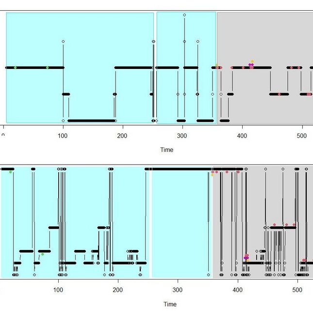
PROJECT: JOINT MODELING WITH HIGH -FREQUENT REPEATED MEASUREMENTS
Data from Kempenhaeghe Centre for Sleep Medicine are the focus of our analysis. In a sleep experiment, the eligible participants with a high risk of heart disturbances were observed for one night of sleep. A multidisciplinary team of researchers (clinical experts, data scientists, engineers) are investigating the effect of sleep characteristics (e.g. body positions and sleep stage) on the occurrence of any of 12 types of arrhythmias obtained from ECG data.
They will implement different analysis approaches. A first analysis is a generalized linear mixed effects model to obtain the relation between body position and the number of heart rhythm disturbances to determine if one position is most likely to generate events. To better capture the time dynamics of the involved variables we will perform various statistical analyses. Joint models will be used to link the various sleep characteristics to the occurrence of each type of event, where we will investigate how each event may lead to a switch of body position and/or how it triggers another event. Given the high frequency of the measurements and the large volume of the available data, we will combine the statistical approaches with machine learning techniques and feature selection techniques to reduce the number of repeated measurements.
The figure Shows profiles of body position and sleep stage for one patient. This patient experienced 6 different types of heart disturbances. The blue area denotes a period with atrial fibrillation and the grey area denotes a period of sinus rhythm deviations. The red dots represent premature atrial contraction, the green dots represents premature ventricular contraction, the yellow dots represent junctional beat, and the purple dots represent fusion beat.