Article

Influence of Environmental and Anthropogenic Factors on Microbial Ecology and Sanitary Threat in the Final Stretch of the Brda River

1
Department of Microbiology, Institute of Experimental Biology, Faculty of Natural Science, Kazimierz Wielki University, 85-090 Bydgoszcz, Poland
2
Department of Experimental Biology, Pomeranian University in Słupsk, 76-200 Słupsk, Poland
*
Author to whom correspondence should be addressed.
Water 2019, 11(5), 922; https://doi.org/10.3390/w11050922
Submission received: 8 April 2019 / Revised: 25 April 2019 / Accepted: 29 April 2019 / Published: 1 May 2019
(This article belongs to the Section Water Quality and Contamination)

Abstract

:
Ecology of aquatic microorganisms depends on a number of environmental parameters. The additional influence of anthropogenic factors is connected with sanitary risk, particularly in urban areas. The study was aimed at assessing the impact of physicochemical and biological parameters on the abundance and activity of bacterioplankton under different spatio-temporal conditions in the urbanized section of the Brda River. The evaluation of sanitary pollution of water was accompanied by the assessment of antibiotic resistance of isolated faecal strains determined using the disk diffusion method. The results indicate that the location of sampling sites significantly affected by the distribution of faecal Escherichia coli and enterococci in the studied part of the river. On the other hand, there were no significant seasonal changes in respiratory activity or abundance of planktonic bacteria. In addition, the abundance of bacterioplankton was not correlated with all measured physico-chemical parameters, though it was correlated with the organic carbon oxidation rate. Depending on the sampling site, bacterial cells with damaged membranes constituted between 8% and 20% of the population. Antibiograms showed the absence of multi-drug resistant strains. Enterococci exhibited the highest resistance to imipenem (45%), while Escherichia coli, to cefoxitin (31%).

1. Introduction

Bacteria play a key role in aquatic ecosystems. As reducers, they participate in the decomposition and circulation of organic matter [1]. The ecological significance of these processes has encouraged investigation into the activity of bacterial communities in water [2,3,4,5]. The results of many studies indicate the lack of a simple relationship between environmental parameters and bacterial populations. Their quantitative and metabolic potential is determined by various biotic and abiotic factors such as trophic state and temperature [6,7]. Moreover, human activities in the catchment area and along shorelines may also affect aquatic ecosystems [8]. Research on the ecology of aquatic microorganisms is additionally impeded by the fact that only a small percentage of these communities can be identified using classical culture methods [9]. In response to stress factors, many bacterial species have developed the ability to enter into a state in which they remain physiologically alive but do not grow or divide (viable but non-culturable state–VBNC). This feature, observed in many pathogens, may increase a potential threat to public health [10]. Bacteria in VBNC state exhibit low metabolic activity [11]. This characteristic is particularly important considering the role that bacteria play in the destruction and transformation of organic matter in aquatic ecosystems. For this reason, the research on bacteria isolated from natural environments should be conducted using modern fluorescent-based methods, which facilitate not only the in situ detection of even very small microorganisms but also the differentiation between viable and non-viable cells. Determining the quantitative structure of bacterioplankton helps understand the role that microorganisms play in decomposition of organic matter, because only viable bacteria participate in matter circulation [12].
In recent years the widespread use of antibiotics in medicine and agriculture has led to the increased occurrence of antibiotic-resistant bacteria in aquatic environments [13,14]. This phenomenon may have serious consequences for public health. In addition, an increased concentration of antibiotic substances in aquatic ecosystems may affect not only the quantitative and qualitative structure of bacteriocenoses but also their ecological role [15]. Taking the above into account we attempted to assess the abundance and activity of the bacterioplankton in the final stretch of the River Brda in an urban area. Additionally, we evaluated sanitary conditions of the studied river section as well as antibiotic resistance of isolated faecal strains. Correlation analyses were conducted to determine factors that influenced the abundance and activity of planktonic bacteria. The hypothesis assumed that the respiratory activity and abundance of bacterioplankton (including indicator bacteria) would not change with the river course in the studied part of the city.

2. Materials and Methods
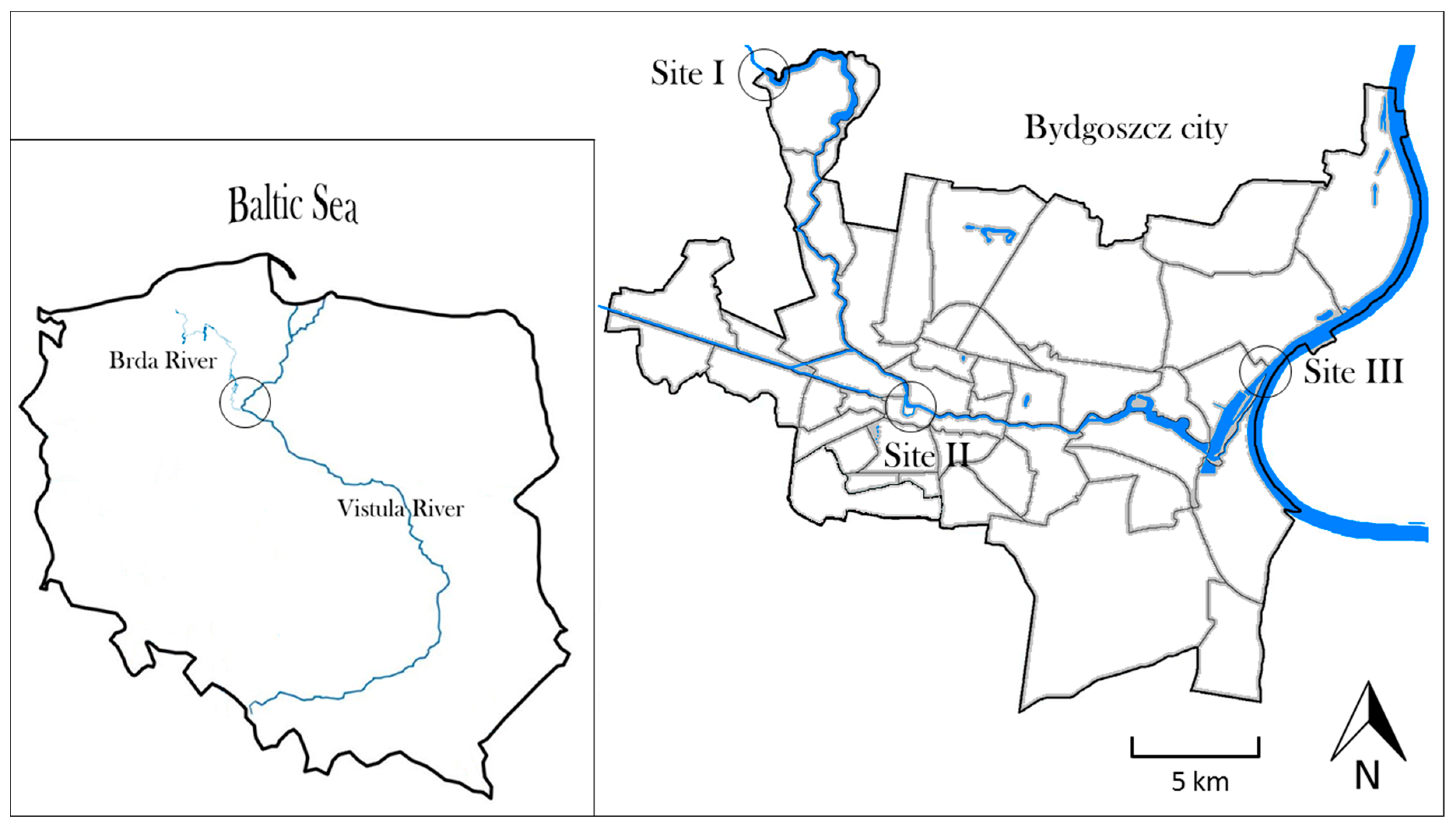
2.1. Study Area

The Brda, a river in northern Poland, is a left tributary of the Vistula, the longest Polish river, which flows into the Baltic Sea. The Brda has the total length of about 240 km and a catchment area of 4665.0 km2. It runs almost entirely through forests. The last stretch of the river, flowing through the city of Bydgoszcz, has numerous recreational and tourist functions [16]. Bydgoszcz population is about 355,000, and the population density is 2017 people per km2.

2.2. Sampling Strategy

Three sampling sites were located within the studied section of the river: 1/on the north-western border of Bydgoszcz, just before the river enters the city, 2/in the centre of Bydgoszcz, where the river is used for recreation, 3/in the north-eastern part of Bydgoszcz, just before its confluence with the Vistula River. The location of sampling sites is shown in Figure 1. Sampling was conducted in a seasonal cycle: in spring (15 May 2017), summer (28 August 2017), and autumn (6 November 2017). Water from the mid-point of the river, depth of about 0.5 m, was collected in triplicate to sterile bottles. The samples were immediately transported to the laboratory and analysed.

2.3. Physico-Chemical Parameters of Water

Prior to microbiological tests, in situ physical and chemical parameters of sampled water, such as temperaturę (T), electrolytic conductivity (EC), pH and oxygen concentration (OC), were determined using Mettler ToledoTM measuring probes (S20 SevenEasys. SG2 SevenGo and FG4 FiveGo). The parameters and the location of sampling sites are presented in Table 1.

2.4. Abundance of Planktonic Bacteria

Classical culture methods and fluorescence-based direct counts were used for quantitative analysis of bacterioplankton. The abundance of heterotrophic bacteria was determined using standard nutrient agar. The quantitative determination of mesophilic bacteria (including faecal bacteria) is particularly important due to the role they play as a faecal pollution marker. Prior to inoculation, water samples were diluted in sterile peptone saline (0.85% aqueous NaCl solution with 1 g of peptone/dm3). Next, 0.1cm3 of each sample was inoculated on sterile nutrient agar (three parallel repetitions). Psychrophilic bacteria were incubated at 20 ± 1 °C for 72 h and mesophilic bacteria, at 35 ± 1 °C for 24 h. Subsequently, all grown colonies were counted and the results were expressed as CFU cm−3 of water.
The abundance of bacterioplankton in water samples was assessed using the LIVE/DEAD® BacLight™ Bacterial Viability Kit. One cm3 of each water sample was transferred to sterile Eppendorf tubes and labeled with 0.15 cm3 propidium iodide (PI) and SYTO®9 in a 1:1 ratio. Samples were incubated for 15 min in the dark at room temperature and filtered through 0.2 μm black membrane filters. The filters were washed with sterile water and placed on slides after drying. With the use of the immersion technique, the slides were immediately viewed under Nikon Eclipse E600 epifluorescence microscope with B-2A filter set (EX 450–490 nm, DM 505 nm, BA 520 nm). The images were archived using Lucia G software. The abundance of bacterioplankton was determined using the following formula:
X = SN/sVR
where:
  • X—number of bacterial cells per 1cm3 of water
  • N—number of cells in a field of view
  • S—effective area of filtration
  • s—analyzed area of the field of view
  • V—volume of filtered sample
  • R—sample dilution
The applied method allowed us to identify cells with damaged and undamaged cell membrane. We calculated the percentage of bacteria with integral cell membrane [MEM+] (stained green with SYTO®9 fluorochrome) and with damaged cell membrane [MEM-] (stained orange with PI marker).

2.5. Distribution of Faecal Bacteria

Escherichia coli were detected using the guidelines of the European standard ISO 9308-1: 2014 [17] and enterococci, ISO 7899-2: 2000 [18]. Escherichia coli were isolated from ChromoCult® Coliform Agar (CCA) selective medium, while faecal streptococci, from Slanetz and Bartley (SB). The selected volume of the water sample was filtered through white filters with 47 mm diameter and 0.45 μm pore size. Plates with filters were incubated for 24 h at 35 ± 1 °C. The number of all characteristic colonies was expressed as CFU 100cm−3 of water.

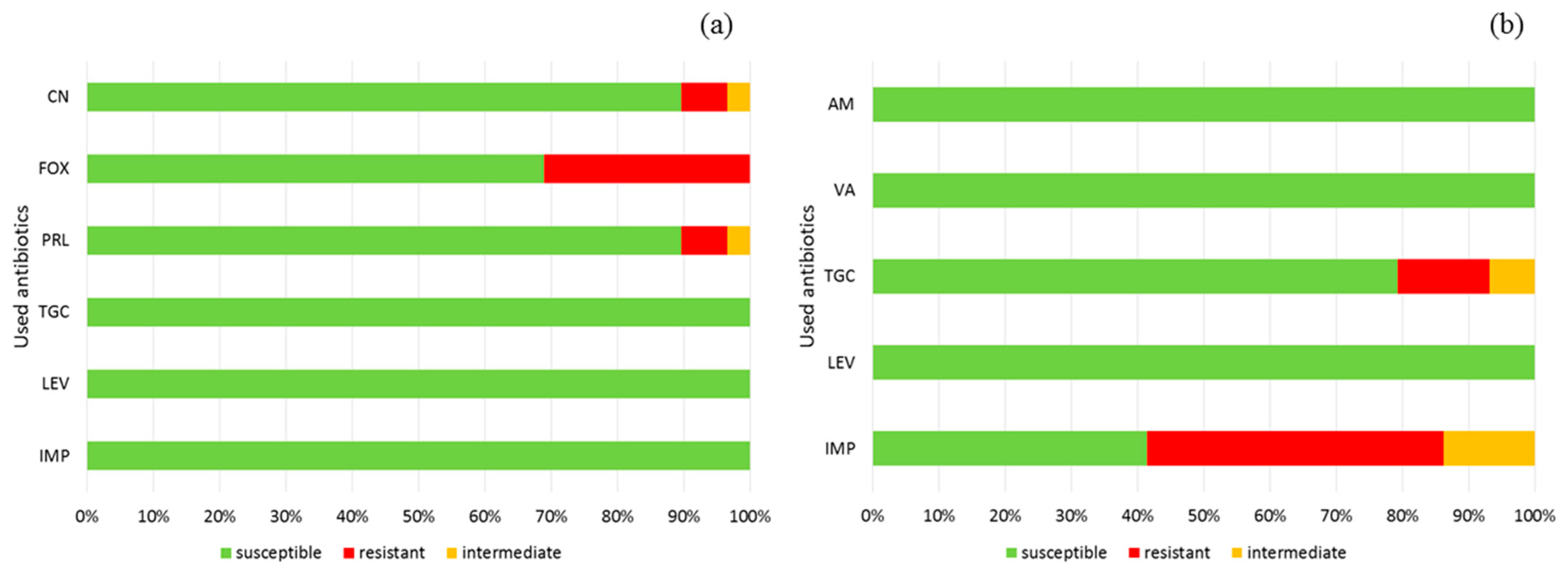
2.6. Antimicrobial Resistance

Antimicrobial resistance of faecal bacteria Escherichia coli isolated from CCA medium and of enterococci isolated from SB medium was determined using the disk diffusion method. The taxonomy of used isolates were confirmed by API® 20E and API® 20 Strep strip tests (bioMérieux, France). Mueller-Hinton Agar was inoculated with bacterial suspension equivalent to 0.5 McFarland standard. Next, paper disks soaked with selected antibiotics were placed on the Petri dishes. After 18 h of incubation at 35 ± 1 °C zones of inhibited growth were measured and the results were compared with the guidelines of The European Committee on Antimicrobial Susceptibility Testing [19]. Information on the antibiotics used in the test can be found in Table 2.

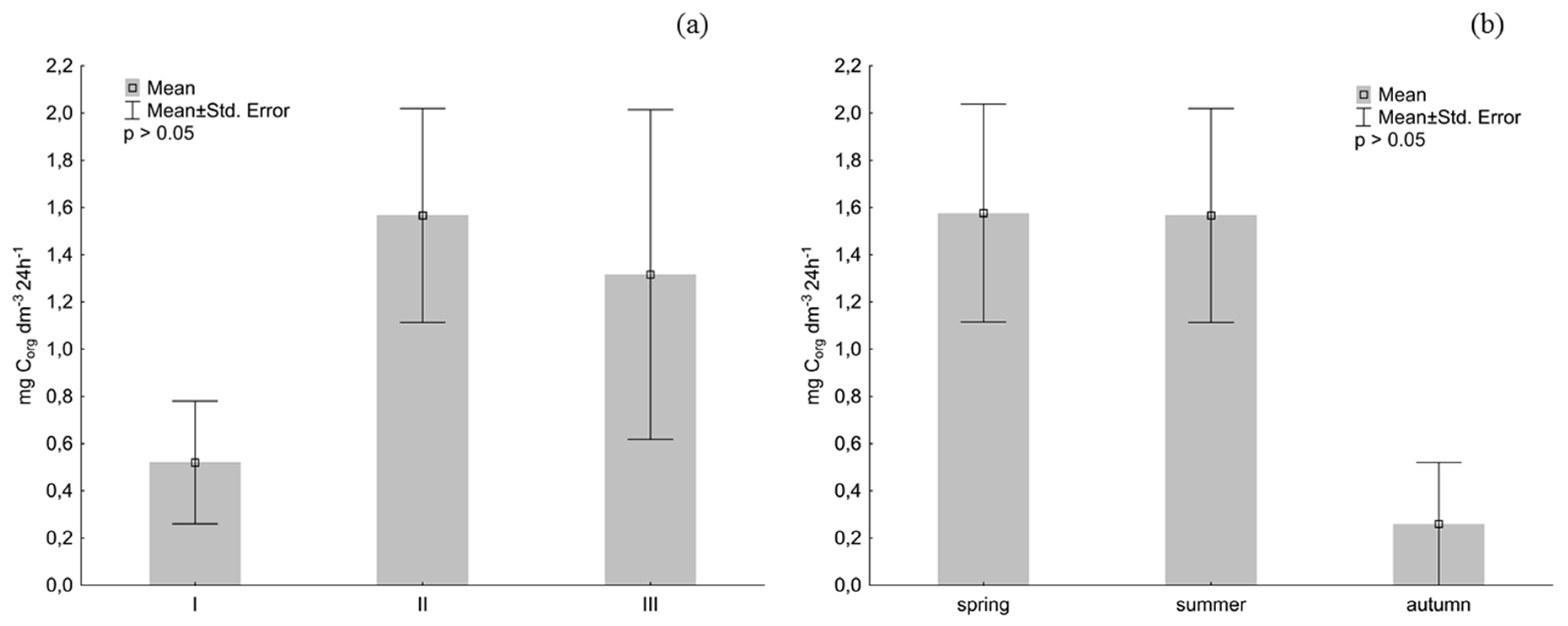
2.7. The Rate of Organic Carbon Oxidation by the Planktonic Bacteria

The rate of organic carbon oxidation was calculated using the respirometric method and OxiTop® Control system (WTW, Poland). The method is based on the measurement of oxygen consumption at a constant temperature, which is accompanied by a pressure change in a sealed sample. Differences between oxygen consumption and carbon dioxide production resulting from the respiratory activity of bacteria are measured by OxiTop-C heads. The OxiTop OC 110 remote controller collects and interprets data using the built-in algorithm. To determine the in situ respiratory activity of planktonic bacteria, 250 cm3 of river water from each sampling site was put into dark OxiTop bottles with a capacity of 500 cm3. Each assay was performed in triplicate. 5 drops of NTH 600 nitrification inhibitor and a magnetic stirrer were placed in the bottles. After a rubber quiver with two tablets (about 0.4 g) of sodium hydroxide (carbon dioxide absorber) was placed in each bottleneck, OxiTop-C measuring heads were tightly screwed on. The parameters were set on the controller, the bottles were placed on a magnetic stirrer and incubated for 24 h at 20 ± 1 °C. The rate of oxygen consumption was used to calculate the rate of carbon mineralization using a 0.29 conversion factor [20]. The results are expressed as mg of oxidized Corg dm3 24 h−1.

2.8. Statistical Analysis
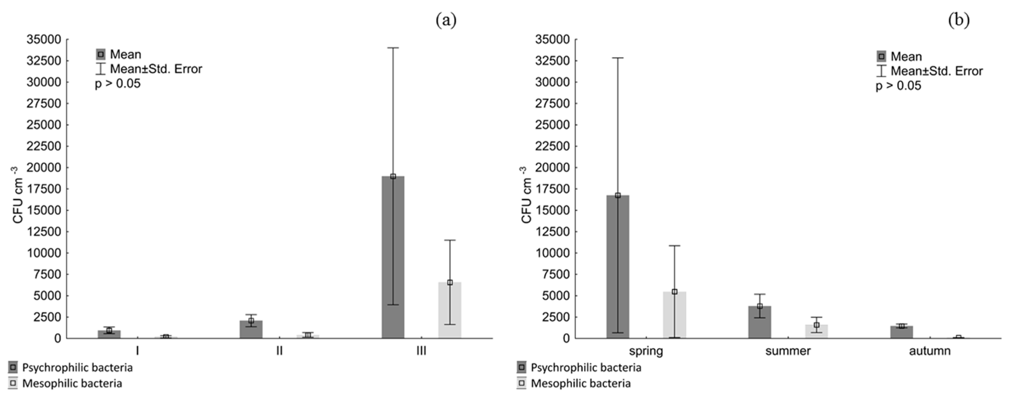
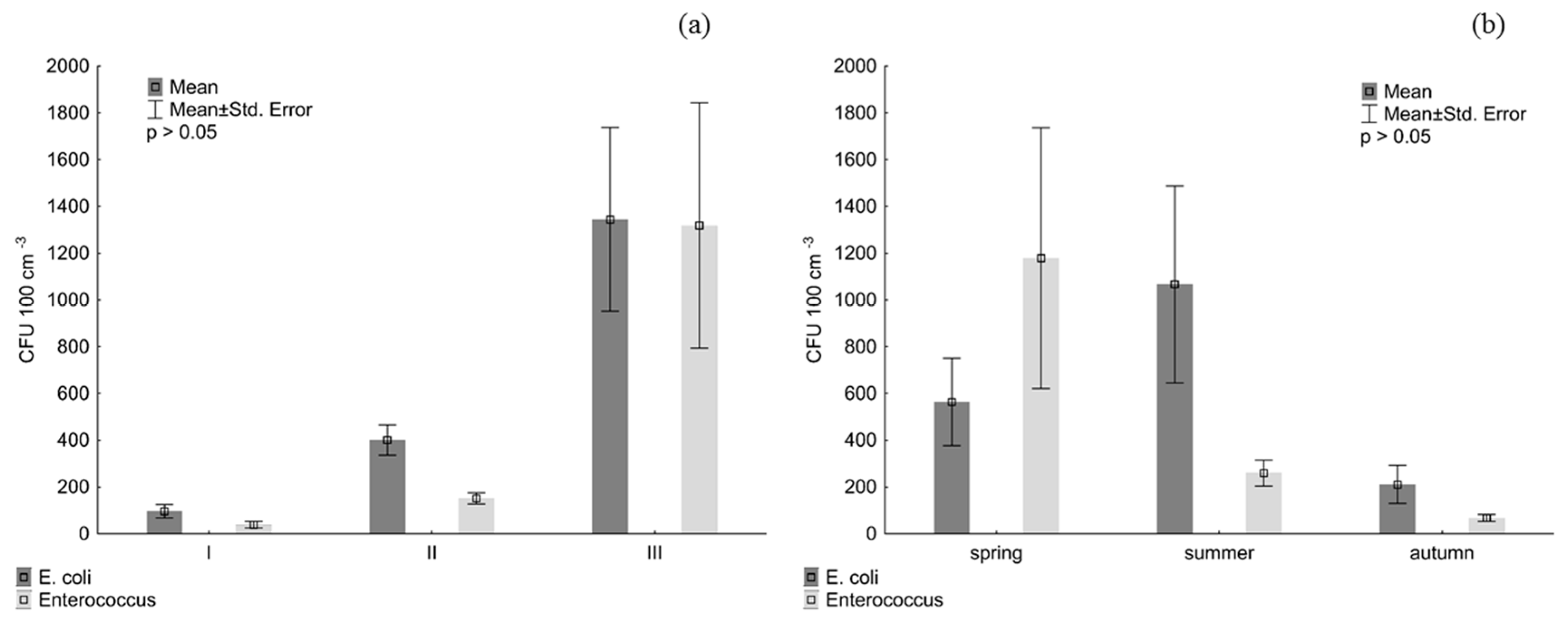
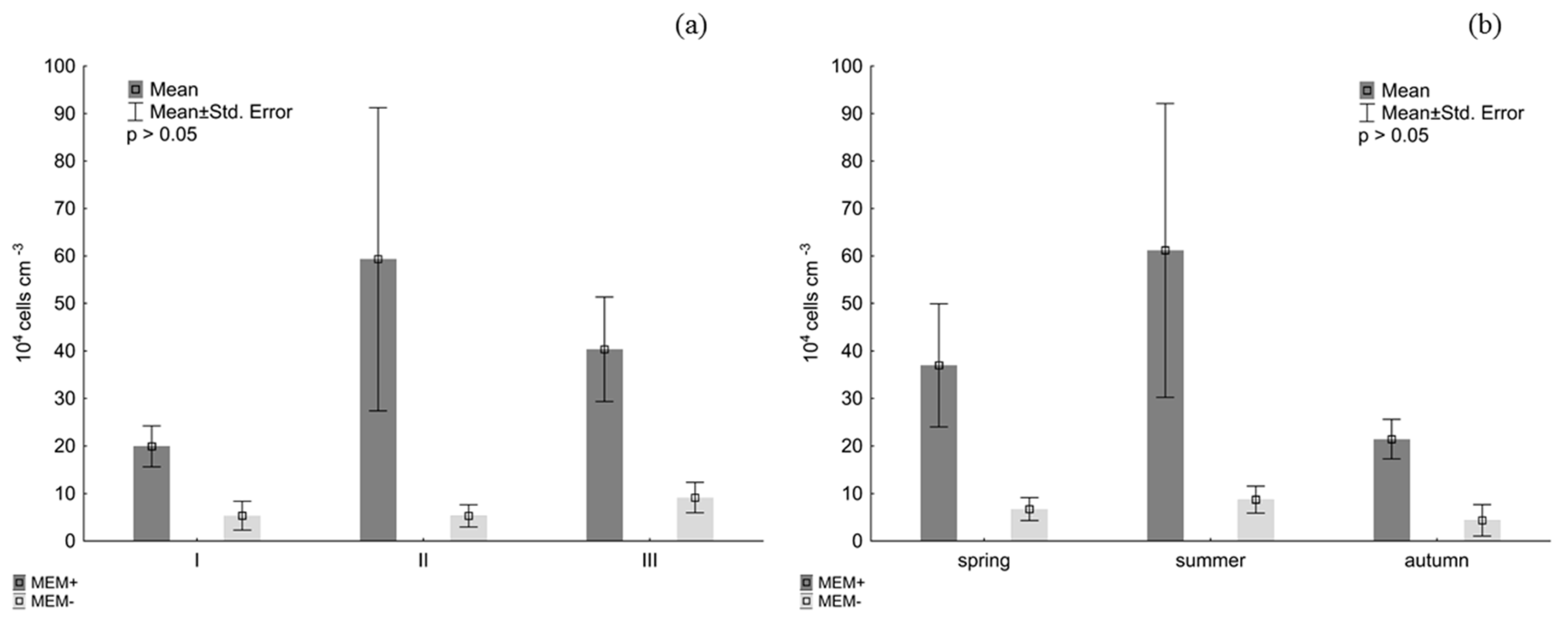
Statistical analyses were conducted using Statistica 13.1 software. The analysis of intergroup differences was based on the Kruskal-Wallis non-parametric test. Post-hoc tests were used to determine intra-group differences. Relationships between the studied parameters were determined using Pearson correlation coefficient. All statistical tests had a significance level p ≤ 0.05.

3. Results

The measurements with classical culture methods indicated that bacterial abundance depended on the location of sampling sites and increased downstream within the studied river section. The lowest abundances of psychrophilic, mesophilic, and indicator bacteria (Escherichia coli and enterococci) were recorded at sampling site I, where the Brda enters the city. In the middle of the investigated river stretch (site II) bacterial abundances were higher, and reached maximum values at sampling site III at the confluence with the Vistula, after the Brda passed through the urban area. (Figure 2 and Figure 3). Statistical analysis showed that the location of sampling sites significantly affected only the abundance of indicator bacteria. Post-hoc tests revealed the largest statistically significant differences between bacterial counts between sites I and III (Table 3). Seasons did not have a significant effect on the abundance of studied bacterial groups, although they were the most abundant in spring and the least abundant in autumn. Only Escherichia coli were the most abundant in summer.
Fluorescence-based direct counts brought slightly different results. Statistical analysis showed that neither the location of sampling sites nor time of the year had a significant impact on the abundance of planktonic bacteria: the highest was recorded at site II in the city centre (5.93 × 10⁵ cells cm−3). The results indicated different seasonal variations in bacterioplankton abundance than previously: the highest was always recorded in summer (6.12 × 10⁵ cells cm−3). The average abundance of planktonic bacteria measured with the use of the fluorescence-based method was one to three orders of magnitude higher than measured with the use of classical culture methods. Cells with damaged membranes constituted between 8.23% (sampling site II) and 20% of the bacterial population (sampling sites I and III) (Figure 4).
As can be seen in Figure 5, organic carbon (Corg) oxidation rate measured with the respirometric method was the lowest at site I (0.52 mg dm−3 24 h−1) and highest at site II (1.57 mg dm−3 24 h−1). Low respiratory activity of bacterioplankton was recorded in autumn, when the average Corg oxidation rate within one day was only 0.26 mg dm−3.
Correlation analysis of the evaluated parameters showed a statistically significant relationship between the organic carbon oxidation rate and dissolved oxygen concentration in water (r = 0.8, p = 0.01). Moreover, respiratory activity of bacterioplankton (expressed as the organic carbon oxidation rate) was significantly correlated with bacterial abundance (r = 0.76, p = 0.02). In the majority of cases, there was a positive correlation between the abundance of the investigated microbial groups and water physicochemical parameters, such as temperature, electrolytic conductivity, and oxygen concentration. A negative correlation was observed only between water pH and the abundance of all studied microbial groups, particularly Escherichia coli and abundance of bacteria. However, the values were not statistically significant (p < 0.05). (Table 4).
Figure 6 demonstrates that the majority of faecal strains were sensitive to the applied antibiotics. enterococci exhibited 100% sensitivity to two of the five tested drugs and high resistance to imipenem, beta-lactam antibiotic of the carbapenem class. About 45% of all isolates were highly resistant to this drug, and about 14%, medium-resistant. Escherichia coli exhibited resistance to two of the six applied antibiotics: approximately 31% of the strains were resistant to cefoxitin, and 7%, to gentamicin and piperacillin.

4. Discussion

Rivers and other aquatic ecosystems play a significant role in the global system of biogeochemical cycles, primarily in the coal cycle [21]. Bacteria are a key element in the circulation of natural elements in these ecosystems. However, little is known about how specific ecological parameters affect bacterial populations [22]. Diversity and abundance of microorganisms determine quality and health of aquatic ecosystems by impacting ecological processes in these environments [23]. This impact, however, may change when large amounts of allochthonous compounds enter their habitat [24]. This is common in urban areas, characterised by a large density of human structures and activities. Since it makes the presence of potentially pathogenic bacteria a real threat, regular monitoring of water quality is highly recommended, especially in recreation areas [25].
In our study, the abundance of studied bacterial groups increased downstream. Particularly significant differences were observed in the distribution of faecal bacteria, the most abundant at the last sampling site, where the total number of Escherichia coli was 1.34 × 10³ CFU 100 cm−3, and of enterococci, 1.32 × 10³ CFU 100cm−3. Although there were no statistically significant seasonal variations, the highest abundances of enterococci, psychrophilic bacteria and mesophilic bacteria were recorded in spring, while the lowest, in autumn. Only Escherichia coli were the most abundant in summer (1.07 × 10³ CFU 100 cm−3). Ma et al. [26] also noted greater spatial than seasonal diversity of river bacterioplankton. The impact of high temperature on bacterial growth was observed only in some sections of the river. Our results did not show any correlation between bacterial abundance and water temperature. It can, therefore, be assumed that an increased number of faecal bacteria in spring and summer was connected with increased participation of Bydgoszcz residents in recreational activities. Donderski et al. [27] maintain that feacal contamination of the Vistula was primarily of human origin. We found that in summer various animals (including birds) build shelters along river banks. Strauch [28] also observed significant seasonal variation in the fluctuations of faecal bacteria in five rivers of the Serengeti National Park in Tanzania. He suggested that animals may be the main contributors to faecal contamination of surface waters, which, in consequence, may pose a serious health threat.
The fluorescence-based method confirmed higher bacterial abundances in summer. Their maxima were recorded at sampling site II, i.e., in the city centre. Sampling location and human influence can determination a bacterial abundance [29]. In our study, statistical analysis did not show any significant differences in spatio-temporal distribution of bacterioplankton. However, the abundance of bacterioplankton measured with this method was several orders of magnitude higher than that measured with classical culture methods. Obviously, this may result from greater accuracy and reliability of the fluorescence-based method but may also indicate that the studied water samples contained bacteria in the VBNC state, in which they remain alive, but cannot divide and grow on culture media. This condition is perceived as cell response to environmental stressors [30]. Human pathogens also possess this ability, which means that although they cannot be grown on conventional media, they retain their virulence [31]. In view of the fact that the presence of VBNC bacteria can pose a serious public health threat, it is recommended that studies on microbial communities should assess the presence of these cells.
As has been already stated, bacterial activity and abundance affect the efficiency of decomposition and transformation of organic matter in the natural environment. The evaluation of bacterial viability using fluorescent dyes is common in medicine, biotechnology, water quality monitoring, and other areas [32]. The method using propidium iodide and SYTO®9 stains demonstrated that bacteria with an integral cell membrane constituted approximately 80% of the bacterial population in the studied water samples. Data presented by other authors suggest that the number of bacteria with undamaged cell membranes depends on the type of ecological niche they inhabit. Perliński et al. [33], examining the share of MEM+ cells in the final stretch of the Słupia River (Ustka, southern Baltic Sea), established that viable bacteria constituted less than 20% of bacterioplankton at all sampling sites. Haglund et al. [34] estimated that viable bacteria constituted 57–63% of the bacteriobenthos in Lake Erken (Sweden). A large contribution of bacteria with an intact cell membrane may indicate an absence of environmental stressors. Another theory states that microbial viability is determined by water pollution and is exihibited by as much as 75% of the bacterial population in contaminated rivers [35].
The evaluation of microbial activity may also be based on the measurement of oxygen consumption using a respirometric technique. Monitoring bacterial respiration is a key element of the research on organic matter transformation in aquatic ecosystems [36]. Our results indicated that the organic carbon oxidation rate was the highest in summer months at sampling site II (city centre). Statistical analysis, however, did not reveal any significant differences in spatio-seasonal variation in the respiratory activity of bacterioplankton. Warkentin et al. [37], studying bacterioplankton in the eutrophic Warnow River (Germany), also recorded the highest bacterial respiration rates in summer (June-August). These results differed significantly from the ones recorded in other months, being even 13 times higher. The authors maintain that bacterial respiration activity was correlated with temperature, but was not correlated with bacterial abundance. Similar observations were made by Zdanowicz et al. [38] studying planktonic and neustonic bacteria in a seaside lake in the Słowiński National Park (Poland). The authors noted that the activity of bacterioplankton was higher than that of bacterioneustone and was susceptible to seasonal effects. They did not find a correlation between bacterial abundance and respiration. Our results indicate that the respiratory activity of planktonic bacteria depended mostly on the concentration of dissolved oxygen in water. In addition, bacterioplankton abundance significantly influenced the organic carbon oxidation rate. Bacterial respiratory activity is considered one of the key factors determining the rate of transformation and mineralization of organic matter in aquatic environments. At the same time, bacterial abundance does not always affect the rate of these processes [39]. The assessment of parameters characterizing the ecology of aquatic microorganisms can be impeded by many factors including changing environmental conditions, different features of investigated water bodies and different research methods. As emphasized by Staley et al. [40], there is a need for a better understanding of spatial and temporal fluctuations in bacterial diversity in aquatic environments. Environmental factors, including physicochemical parameters, should also be carefully analysed.
The WHO report warns that the acquisition of antibiotic resistance by bacteria is becoming increasingly common and may pose a public health threat. At the same time, the document accentuates the need to monitor and control this tendency [41]. A vast majority of studies investigate drug resistance of bacterial strains isolated from sick or healthy patients [42]. Natural environments, where the presence of potential pathogens may pose a sanitary risk, are examined less regularly. The current research show that in aquatic environment occurrence of antibiotic-resistance faecal coliform bacteria with may have a public health implication [43]. This is especially important in ecosystems subjected to a high anthropogenic pressure. The high abundance of bacterial pathogens in water ecosystem can be a potential health threat for the local population [44]. Boon et al. [45] observed that faecal strains isolated from urbanized sections of the river exhibited higher antibiotic resistance than those isolated from non-urbanized parts. The authors also noted that Escherichia coli isolated from all sampling sites exhibited 100% resistance to penicillin. In our study, Escherichia coli exhibited the highest resistance to cefoxitin (31%). Only about 7% of the isolates were resistant to gentamicin form aminoglycosides group and piperacillin from penicillin group. As noticed by Aslan et al. [46] further research is needed to evaluate health risks related to occurrence of antibiotic-resistance bacteria in aquatic environments and using water for public purposes.

5. Conclusions

Our results indicate higher spatial rather than temporal variation of bacterioplankton in the studied section of the Brda River, particularly the abundance of faecal bacteria increased with the river transition to the mouth. The abundance of bacterioplankton was not significantly correlated with water physicochemical parameters but it was correlated with the organic carbon oxidation rate. The antibiotic resistance analysis did not reveal any multi-drug resistant faecal strains. In view of the potential sanitary risk, it is recommended that the research on the microbial ecology of aquatic environments in urban areas should contain the assessment of the distribution of faecal bacteria and of their antimicrobial resistance.

Author Contributions

Ł.K. designed the study, data analysis and the manuscript preparation; M.M-A. and E.J. performed the laboratory analysis; E.D. and P.P. review and editing of the manuscript; K.H. contributed to sample collection.

Funding

This study was supported by the Polish Minister of Science and Higher Education, under the program “Regional Initiative of Excellence” in 2019–2022 (Grant No. 008/RID/2018/19).

Conflicts of Interest

The authors declare no conflict of interest.

References

  1. Newton, R.J.; Jones, S.E.; Eiler, A.; McMahon, K.D.; Bertilsson, S. A Guide to the Natural History of Freshwater Lake Bacteria. Microbiol. Mol. Biol. Rev. 2011, 75, 14–49. [Google Scholar] [CrossRef]
  2. Kirchman, D.L.; Dittel, A.I.; Findlay, S.E.G.; Fischer, D. Changes in bacterial activity and community structure in response to dissolved organic matter in the Hudson River, New York. Aquat. Microb. Ecol. 2004, 35, 243–257. [Google Scholar] [CrossRef] [Green Version]
  3. Kiersztyn, B.; Siuda, W.; Chróst, R.J. Persistence of bacterial proteolytic enzymes in lake ecosystems. FEMS Microb. Ecol. 2012, 80, 124–134. [Google Scholar] [CrossRef] [Green Version]
  4. Krevš, A.; Kučinskienė, A. Influence of invasive Acer negundo leaf litter on benthic microbial abundance and activity in the littoral zone of a temperate river in Lithuania. Knowl. Manag. Aquat. Ecosyst. 2017, 418, 26. [Google Scholar] [CrossRef]
  5. Perliński, P.; Mudryk, Z.J.; Antonowicz, J. Enzymatic activity in the surface microlayer and subsurface water in the harbour channel. Estuar. Coast. Shelf Sci. 2017, 196, 150–158. [Google Scholar] [CrossRef]
  6. Zhang, M.; Yu, N.; Chen, L.; Jiang, C.; Tao, Y.; Zhang, T.; Chen, J.; Xue, D. Structure and se-asonal dynamics of bacterial communities in three urban rivers in China. Aquat. Sci. 2012, 74, 113–120. [Google Scholar] [CrossRef]
  7. Dai, Y.; Yang, Y.; Wu, Z.; Feng, Q.; Xie, S.; Liu, Y. Spatiotemporal variation of planktonic and sediment bacterial assemblages in two plateau freshwater lakes at different trophic status. Appl. Microbiol. Biotechnol. 2016, 100, 4161–4175. [Google Scholar] [CrossRef]
  8. Krevš, A.; Kučinskienė, A. Microbial decomposition of sedimentary organic matter in small temperate lakes. Fundam. Appl. Limnol. 2018, 191, 239–251. [Google Scholar] [CrossRef]
  9. Amann, R.I.; Ludwig, W.; Schleifer, K.H. Phylogenetic identification and in situ detection of individual microbial cells without cultivation. Microbiol. Rev. 1995, 59, 143–169. [Google Scholar]
  10. Oliver, J.D. Recent findings on the viable but nonculturable state in pathogenic bacteria. FEMS Microbiol. Rev. 2010, 34, 415–425. [Google Scholar] [CrossRef] [Green Version]
  11. Oliver, J.D. The viable but nonculturable state in bacteria. J. Microbiol. 2005, 43, 93–100. [Google Scholar]
  12. Senjarini, K.; Karsten, U.; Schumann, R. Application of Fluorescence Markers for the Diagnosis of Bacterial Abundance and Viability in Aquatic Ecosystem. J. Microbiol. Res. 2013, 3, 143–147. [Google Scholar]
  13. Dang, H.; Ren, J.; Song, L.; Sun, S.; An, L. Diverse tetracycline resistant bacteria and resi-stance genes from coastal waters of Jiaozhou Bay. Microb. Ecol. 2008, 55, 237–246. [Google Scholar] [CrossRef] [PubMed]
  14. Ramesh, S.; Manivasagan, S.; Ashokkumar, S.; Rajaram, G.; Mayavu, P. Plasmid profiling and multiple antibiotic resistance of heterotrophic bacteria isolated from Muthupettai mangrove environment, southeast coast of India. Curr. Res. Bacteriol. 2010, 3, 227–237. [Google Scholar] [CrossRef]
  15. Costanzo, S.D.; Murby, J.; Bates, J. Ecosystem response to antibiotic entering the aquatic environment. Mar. Pollut. Bull. 2005, 51, 218–223. [Google Scholar] [CrossRef]
  16. Radtke, G.; Bernaś, R.; Dębowski, P.; Morzuch, J.; Skóra, M. Fish fauna of the Brda river system. Sci. Annu. Pol. Angling Assoc. 2015, 28, 43–84. [Google Scholar]
  17. ISO 9308-1:2014. Water Quality—Enumeration of Escherichia coli and Coliform Bacteria—Part 1 Membrane Filtration Method for Waters with Low Bacterial Background Flora; International Organization for Standardization: Geneva, Switzerland, 2014. [Google Scholar]
  18. ISO 7899-2:2000. Water Quality—Detection and Enumeration of Intestinal Enterococci—Part 2: Membrane Filtration Method; International Organization for Standardization: Geneva, Switzerland, 2000. [Google Scholar]
  19. The European Committee on Antimicrobial Susceptibility Testing. Breakpoint Tables for Interpretation of MICs and Zone Diameters. Version 8.1. 2018. Available online: http://www.eucast.org (accessed on 15 May 2018).
  20. Sepers, A.B.J. The aerobic mineralization of organic compounds in the saline Lake Grevelingen (The Netherlands) (VI). Hydrobiol. Bull. 1979, 13, 81–82. [Google Scholar] [CrossRef]
  21. Aufdenkampe, A.K.; Mayorga, E.; Raymond, P.A.; Melack, J.M.; Doney, S.C.; Alin, S.R.; Aalto, R.E.; Yoo, K. Riverine coupling of biogeochemical cycles between land, oceans, and atmosphere. Front. Ecol. Environ. 2011, 9, 53–60. [Google Scholar] [CrossRef] [Green Version]
  22. Eiler, A.; Heinrich, F.; Bertilsson, S. Coherent dynamics and association networks among lake bacterioplankton taxa. ISME J. 2012, 6, 330–342. [Google Scholar] [CrossRef] [PubMed]
  23. Jeffries, T.C.; Schmitz Fontes, M.L.; Harrison, D.P.; Van-Dongen-Vogels, V.; Eyre, B.D.; Ralph, P.J.; Seymour, J.R. Bacterioplankton Dynamics within a Large Anthropogenically Impacted Urban Estuary. Front. Microbiol. 2016, 6, 1438. [Google Scholar] [CrossRef] [PubMed]
  24. Carney, R.L.; Mitrovic, S.M.; Jeffries, T.; Westhorpe, D.; Curlevski, N.; Seymour, J.R. River bacterioplankton community responses to a high inflow event. Aquat. Microb. Ecol. 2015, 75, 187–205. [Google Scholar] [CrossRef]
  25. Zmysłowska, I.; Gołaś, I. Sanitary and bacteriological examinations of lake Oświn water. Pol. J. Environ. Stud. 2003, 12, 351–356. [Google Scholar]
  26. Ma, L.; Mao, G.; Liu, J.; Gao, G.; Zou, C.; Bartlam, M.G.; Wang, Y. Spatial-Temporal Chan-ges of Bacterioplankton Community along an Exhorheic River. Front. Microbiol. 2016, 7, 250. [Google Scholar] [CrossRef]
  27. Donderski, W.; Wilk, I. The Sanitary State of Water in the River Vistula between Wyszogród and Toruń. Pol. J. Environ. Stud. 2002, 11, 509–515. [Google Scholar]
  28. Strauch, A.M. Seasonal variability in faecal bacteria of semiarid rivers in the Serengeti National Park, Tanzania. Mar. Freshw. Res. 2011, 62, 1191–1200. [Google Scholar] [CrossRef]
  29. Quintela-Alonso, P.; Pérez-Uz, B.; Sanchez-Jimenez, A.; Murciano, A.; Centeno, J.D.; García-Rodríguez, M.; Montero, E.; Muñoz, B.; Olmedo, C.; Refoyo, P.; et al. Complexity of river ciliate communities at a national park highlights the need for microbial conservation. Aquat. Conserv.: Mar. Freshw. Ecosyst. 2018, 28, 408–421. [Google Scholar] [CrossRef]
  30. Lleò, M.M.; Benedetti, D.; Tafi, M.C.; Signoretto, C.; Canepari, P. Inhibition of the resusci-tation from the viable but non-culturable state in Enterococcus faecalis. Environ. Microbiol. 2007, 9, 2313–2320. [Google Scholar] [CrossRef]
  31. Ramamurthy, T.; Ghosh, A.; Pazhani, G.P.; Shinoda, S. Current perspectives on viable but non-culturable (VBNC) pathogenic bacteria. Front. Public Health 2014, 2, 103. [Google Scholar] [CrossRef]
  32. Stiefel, P.; Schmidt-Emrich, S.; Maniura-Weber, K.; Ren, Q. Critical aspects of using bac-terial cell viability assays with the fluorophores SYTO9 and propidium iodide. BMC Microbiol. 2015, 15, 36. [Google Scholar] [CrossRef]
  33. Perliński, P.; Skórczewski, P.; Zdanowicz, M.; Mudryk, Z. Participation of MEM + bacte-ria in the bacterioplankton community in Ustka harbor, the River Słupia estuary, Southern Baltic Sea. Eur. J. Biol. Res. 2015, 5, 17–24. [Google Scholar]
  34. Haglund, A.L.; Lantz, P.; Törnblom, E.; Tranvik, L. Depth distribution of active bacteria and bacterial activity in lake sediment. FEMS Microb. Ecol. 2003, 46, 31–38. [Google Scholar] [CrossRef] [Green Version]
  35. Porter, J.; Diaper, J.; Edwards, C.; Pickup, R. Direct Measurements of Natural Planktonic Bacterial Community Viability by Flow Cytometry. Appl. Environ. Microbiol. 1995, 61, 2783–2786. [Google Scholar]
  36. Swiontek-Brzezinska, M.; Lalke-Porczyk, E.; Donderski, W.; Walczak, M. Occurrence and activity of microorganisms in shrimp waste. Curr. Microbiol. 2008, 57, 580–587. [Google Scholar] [CrossRef]
  37. Warkentin, M.; Freese, H.M.; Schumann, R. Bacterial Activity and Bacterioplankton Diversity in the Eutrophic River Warnow-Direct Measurement of Bacterial Growth Efficiency and Its Effect on Carbon Utilization. Microb. Ecol. 2011, 61, 190–200. [Google Scholar] [CrossRef]
  38. Zdanowicz, M.; Mudryk, Z.J. Abundance, production and respiration of bacterioneu-ston and bacterioplankton in the coastal lake Dołgie Wielkie. Balt. Coast. Zone 2017, 21, 73–86. [Google Scholar]
  39. Kubera, Ł.; Donderski, W. Distribution and activity of benthic bacteria in four lakes in the Bory Tucholskie National Park (Poland). Aquat. Microb. Ecol. 2017, 79, 127–135. [Google Scholar] [CrossRef]
  40. Staley, C.; Gould, T.J.; Wang, P.; Phillips, J.; Cotner, J.B.; Sadowsky, M.J. Species sorting and seasonal dynamics primarily shape bacterial communities in the Upper Mississippi River. Sci. Total Environ. 2015, 505, 435–445. [Google Scholar] [CrossRef]
  41. World Health Organization. Antimicrobial Resistance; Global Report on Surveillance; World Health Organization: Geneva, Switzerland, 2014. [Google Scholar]
  42. Cho, S.H.; Lim, Y.S.; Park, M.S.; Kim, S.H.; Kang, Y.H. Prevalence of antibiotic resistance in Escherichia coli fecal isolates from healthy persons and patients with diarrhea. Osong Public Health Res. Perspect. 2011, 2, 41–45. [Google Scholar] [CrossRef]
  43. Mudryk, Z.J.; Perliński, P.; Robak, D. Antibiotic resistance of fecal coliform bacteria in-habiting sea water and sand of marine recreation beach in the southern Baltic Sea. Balt. Coast. Zone 2016, 20, 25–36. [Google Scholar]
  44. Sepulveda-Jauregui, A.; Hoyos-Santillan, J.; Gutierrez-Mendieta, F.J.; Torres-Alvarado, R.; Dendooven, L.; Thalasso, F. The impact of anthropogenic pollution on limnological characteristics of a subtropical highland reservoir “Lago de Guadalupe”, Mexico. Knowl. Manag. Aquat. Ecosyst. 2013, 410, 04. [Google Scholar] [CrossRef]
  45. Boon, P.I.; Cattanach, M. Antibiotic resistance of native and faecal bacteria isolated from rivers, reservoirs and sewage treatment facilities in Victoria, south-eastern Australia. Lett. Appl. Microbiol. 1999, 28, 164–168. [Google Scholar] [CrossRef] [PubMed] [Green Version]
  46. Aslan, A.; Cole, Z.; Bhattacharya, A.; Oyibo, O. Presence of Antibiotic-Resistant Escherichia coli in Wastewater Treatment Plant Effluents Utilized as Water Reuse for Irrigation. Water 2018, 10, 805. [Google Scholar] [CrossRef]
Figure 1. Arrangement of the sampling sites. The rivers are marked in blue.
Water 11 00922 g001
Figure 2. Average number of heterotrophic bacteria (a) at the research sites, (b) in the research seasons.
Water 11 00922 g002
Figure 3. Average number of faecal indicator bacteria (a) at the research sites, (b) in the research seasons.
Water 11 00922 g003
Figure 4. Total abundance of planktonic bacteria with the integral cell membrane (MEM+) and damaged cell membrane (MEM-) (a) at the research sites, (b) in the research seasons.
Water 11 00922 g004
Figure 5. Rate of organic carbon oxidation by planktonic bacteria (a) at the research sites, (b) in the research seasons.
Water 11 00922 g005
Figure 6. Antimicrobial resistance of isolated strains (n = 29) for Enterococcus (a) and Escherichia coli (b). AM–Ampicillin, CN– Gentamicin, FOX–Cefoxitin, IMP–Imipenem, LEV–Levofloxacin, PRL–Piperacillin, TGC–Tigecycline, VA–Vancomycin.
Water 11 00922 g006
Table 1. Physico-chemical parameters of water. Results are presented as means ± SD; range is given in brackets.
SiteLocationT (°C)pHEC (µS cm−1)OC (mg dm−3)
I53°12′16,86″ N15.7 ± 5.667.6 ± 0.58350.4 ± 36.935.4 ± 0.96
17°56′20,19″ E(8.8–22.1)(6.6–8.0)(303.0–390.0)(4.1–5.7)
II53°7′25,48″ N15.0 ± 5.027.7 ± 0.45360.1 ± 21.715.8 ± 0.83
17°59′53,81″ E(8.6–20.8)(7.0–8.0)(331.0–382.0)(4.6–6.5)
III53°7′22,28″ N14.8 ± 4.927.5 ± 0.32367.7 ± 20.925.7 ± 0.7
18°5′14,65″ E(8.7–20.7)(7.1–7.9)(339.0–397)(4.8–6.5)
T–temperature, EC–electrolytic conductivity, OC–oxygen concentration.
Table 2. Characteristic of used antibiotics.
Class of AntibioticType of AntibioticSymbol of DiscConcentration (µg)
CarbapenemsImipenemIMP10
FluoroquinolonesLevofloxacinLEV5
TetracyclinesTigecyclineTGC15
GlycopeptidesVancomycin *VA5
PenicillinsAmpicillin *AM2
Piperacillin **PRL30
CephalosporinsCefoxitin **FOX30
AminoglycosidesGentamicin **CN10
* Only for Enterococcus spp.; ** Only for Escherichia coli.
Table 3. Statistical differences in the number of indicator bacteria between the analyzed datasets based on post-hoc for Kruskal-Wallis test.
Escherichia coliEnterococcus spp.
IIIIIIIIIIII
I
II* *
III**ns ***ns
*p < 0.05; **p < 0.01; ***p < 0.001, ns: not significant.
Table 4. Correlations between physico-chemical and biological parameters of water and the number of analyzed group of bacteria.
T (°C)pHEC (μS cm−1)OC (mg dm−3)Corg (dm3 24 h−1)
Correlation (r)
Psychrophilic bacteria0.17−0.010.240.380.57
Mesophilic bacteria0.21−0.050.210.40.59
Escherichia col0.33−0.34−0.080.220.48
Enterococcus spp.0.18−0.010.240.40.59
Abundance of bacteria0.44−0.47−0.310.50.76 *
Corg (dm3 24 h−1)0.67−0.35−0.210.8*
T–temperature, EC–electrolytic conductivity, OC–oxygen concentration; * p < 0.05.

