Detection of Infectious Noroviruses from Wastewater and Seawater Using PEMAXTM Treatment Combined with RT-qPCR


Institute of Environmental Science and Research Ltd. (ESR), Porirua 5240, New Zealand
*
Author to whom correspondence should be addressed.
Water 2018, 10(7), 841; https://doi.org/10.3390/w10070841
Submission received: 19 May 2018
/
Revised: 14 June 2018
/
Accepted: 22 June 2018
/
Published: 25 June 2018
(This article belongs to the Special Issue Health Risks of Alternative Water Sources)
Abstract
:Rapid detection of infectious noroviruses from environmental samples is essential to minimize the risk of norovirus outbreaks associated with environmental transmission. Reverse transcription quantitative real-time polymerase chain reaction (RT-qPCR) methods are rapid and sensitive, but cannot differentiate between infectious and non-infectious noroviruses. In this study, a PEMAXTM treatment followed by RT-qPCR (PEMAXTM-RT-qPCR) method was developed for murine norovirus and norovirus GI/GII, and evaluated for the selective detection of infectious viruses following heat inactivation. The norovirus PEMAXTM-RT-qPCR method was then evaluated for the selective detection of infectious viruses from environmental samples. Following heat-treatment (90 °C for 3 min), the murine norovirus PEMAXTM-RT-qPCR showed at least a 2.04 log10 reduction in detectable virus, compared to a 0.43 log10 reduction for RT-qPCR alone. Under the same conditions, the norovirus PEMAXTM-RT-qPCR showed a 0.34 to 0.98 log10 (GI.3) and 0.63 to 2.06 log10 (GII.4) reduction in detectable viruses, compared to 0.05 to 0.18 log10 (GI.3) and 0.06 to 0.25 log10 (GII.4) for RT-qPCR alone. Evaluation of the norovirus PEMAXTM-RT-qPCR on norovirus-contaminated influent and effluent wastewater, and seawater indicated a high proportion of non-infectious norovirus GI and GII (i.e., 56 to 100% in seawater, 32 to 76% in effluent, and 11 to 79% in influent) was present in samples. While potentially overestimating the amount of infectious noroviruses, this approach has potential to provide better information on viral infectivity than RT-qPCR alone.
1. Introduction
Understanding persistence and inactivation of norovirus in the environment has been limited due to the lack of a robust, reproducible, and quantitative detection method that is able to detect only infectious viruses. Currently, sensitive and specific reverse transcription quantitative real-time polymerase chain reaction (RT-qPCR) methods are widely used for the detection and quantification of noroviruses from a wide variety of samples including water and food [1,2]. A disadvantage of this approach is that a positive RT-qPCR result can be produced by non-infectious viruses present in the sample [3], and therefore RT-qPCR data may not be suitable for a health risk assessment.
A viral receptor binding approach utilizing histo-blood group antigens has been used with the aim of detecting infectious noroviruses, however, the process is expensive, labor intensive, and not suitable for all norovirus strains [4,5]. Recently an in vitro norovirus culture method was successfully developed [6,7] but it is not straightforward to implement, expensive, and currently limited to a few research laboratories worldwide. Therefore, other approaches for the detection of infectious noroviruses from environmental samples are needed.
Photoactivatable dyes such as propidium monoazide (PMA) and ethidium monoazide (EMA) (Biotium, Inc., Fremont, CA, USA) have been used with RT-qPCR with some success for the selective detection of infectious norovirus using RT-qPCR [8,9,10,11,12,13]. Theoretically, these dyes penetrate a damaged capsid and, in the presence of light, a covalent bond forms with the nucleic acid, resulting in a reduction in nucleic acid extraction efficiency and in the RT-qPCR signal [14]. In studies with bacteria, Nocker et al. [15] reported that PMA did not enter inactivated bacteria with an intact cell membrane, and consequently overestimated their infectivity. Conversely, EMA entered the intact cell membrane of viable bacteria and underestimated their infectivity. A study on the combined use of PMA and EMA for the detection of Salmonella Enteriditis found that this approach was more effective for discriminating non-viable bacteria from viable ones than PMA and EMA alone [16]. An alternative approach is the double photoactivatable dye PEMAXTM (GenIUL, Barcelona, Spain), which consists of both PMA and EMA. To our knowledge, one study has evaluated the performance of PEMAXTM for the detection of infectious noroviruses, but the study lacked optimization of the PEMAXTM concentration [9].
In our study, the concentration of PEMAXTM for the selective detection of infectious viruses was optimized, and the efficiency of PEMAXTM-RT-qPCR evaluated for the selective detection of infectious murine norovirus and norovirus GI.3 and GII.4 from heat-inactivated samples. The newly developed PEMAXTM-RT-qPCR method was further evaluated using norovirus-contaminated influent and effluent wastewater and seawater samples.
2. Materials and Methodology
2.1. Viral Stock Suspensions
Murine norovirus was propagated in RAW 264.7 mouse leukemic monocyte/macrophage cells and quantified using a monolayer plaque assay to determine the plaque forming units (PFU)/mL as described elsewhere [17,18] and stored at −80 °C until required. The murine norovirus was diluted to give a 105 PFU/mL stock suspension.
Clarified suspensions (10% (wt/vol) in viral transport media) of norovirus GI.3 and GII.4 positive human faecal samples, submitted to ESR for norovirus outbreak surveillance purposes, were prepared and the stock suspensions stored at 5 °C until required (~4 weeks).
2.2. Recovery of Noroviruses from Wastewater and Seawater
Archived virus concentrations from wastewater and seawater samples were used in this study. Briefly, viruses from influent wastewater (2 L) collected from six different wastewater treatment plants were concentrated using beef extract elution followed by polyethylene glycol 6000 precipitation (10% wt/vol) with the addition of sodium chloride (1.75% wt/vol) [19]. Viruses from effluent wastewater (10 L) collected from two treatment plants and seawater (10 L) collected from an estuarine harbor were concentrated using an ultrafiltration method, and further concentrated using polyethylene glycol 6000 and sodium chloride as above [1]. Final concentrates of between 5–10 mL were obtained and stored at −80 °C until required.
2.3. Heat Inactivation of Murine Norovirus and Norovirus
To achieve inactivation of murine norovirus and norovirus GI.3 and GII.4 for the method development and evaluation, 100 μL aliquots of each virus stock suspension were transferred into 200 μL PCR tubes and heated at 90 °C for 3 min using a BioRad CFX thermal cycler (Bio-Rad Laboratories, Hercules, CA, USA). Two 100 μL aliquots were pooled to give 200 μL volumes for subsequent use.
2.4. Optimisation of PEMAXTM Concentration
To optimize the PEMAXTM (GenIUL) concentration, 0.5 mg PEMAXTM was first dissolved in 500 µL dimethyl sulfoxide (20% vol/vol) (Sigma-Aldrich, Auckland, New Zealand) to obtain a 2 mM working solution. Aliquots (200 µL) of heat-inactivated and non-heat-inactivated murine norovirus was transferred into 1.5 mL transparent centrifuge tube (GenIUL), and either 0, 50, 100, or 200 µM PEMAXTM solution added to give a total volume of up to 225 µL. The tubes were then incubated in the dark for 30 min and treated with the “Photo Activation System for Tubes” PhAST blue light (GenIUL) for 15 min. Viral nucleic acid was then extracted from the entire sample (at least 200 µL) and tested for murine norovirus using a one-step RT-qPCR method (Section 2.8). Triplicate samples were processed to ensure the reproducibility of the method.
2.5. Evaluation of PEMAXTM-RT-qPCR for the Selective Detection of Infectious Murine Norovirus
An initial experiment was conducted to evaluate the efficiency of the PEMAXTM-RT-qPCR method for the selective detection of infectious murine norovirus. Ten-fold serial dilutions (10−1 to 10−4) of murine norovirus stock suspension (105 PFU/mL) were prepared and divided into four (200 µL) aliquots. Two sets of aliquots of 100 to 10−4 dilutions were heat-inactivated (90 °C for 3 min). One set of aliquots was treated with the optimal concentration of PEMAXTM (Section 2.4), and another set used as a control. Two sets of aliquots of 100 to 10−4 dilutions were used to determine the percentage of infectious viruses in the stock suspension. This was achieved by treating one set of original (non-heat-inactivated) aliquots with PEMAXTM and the other remained untreated. Viral nucleic acid was extracted from the entire sample (at least 200 µL) and tested for murine norovirus using a one-step RT-qPCR method (Section 2.8).
2.6. Evaluation of PEMAXTM-RT-qPCR for the Selective Detection of Infectious Norovirus
As above, experiments were conducted to determine the efficiency of the PEMAXTM-RT-qPCR method for the selective detection of infectious norovirus GI.3 and GII.4. Ten-fold serial dilutions (10−1 to 10−4) of each genotype were prepared from the stock suspension (Section 2.1) and treated with PEMAXTM (Section 2.4). Viral nucleic acid was extracted from the entire sample (at least 200 µL) and tested for norovirus GI and GII using a duplex one-step RT-qPCR method (Section 2.8).
2.7. Application of PEMAXTM-RT-qPCR for Detection of Infectious Norovirus from Environmental Samples
Two 200 µL aliquots of each wastewater and seawater concentrate were prepared for each experiment. One aliquot was treated with PEMAXTM at the optimized concentration (Section 2.4) and the other remained untreated. Viral nucleic acid was then extracted from the entire sample (at least 200 µL) and tested using a duplex one-step RT-qPCR method (Section 2.8).
2.8. Nucleic Acid Extraction and Virus Detection
Prior to analysis by RT-qPCR, viral nucleic acid was extracted from 200–225 µL (depending on the volume) of each sample using the High Pure Viral Nucleic Acid Kit (Roche Life Science, Mannheim, Germany) according to the manufacturer’s instructions. A reagent blank (water) was included during viral nucleic acid extraction. All nucleic acid samples were tested for potential PCR inhibition by using a sketa22 qPCR method as described elsewhere [20]. No PCR inhibition was detected in any of the samples and were used for downstream analysis.
Previously published primers and probes were used for the detection of norovirus GI/GII [21] and murine norovirus [18]. One-step RT-qPCR was performed using the SuperScriptTM III PlatinumTM One-Step RT-qPCR Kit (Invitrogen Corporation, Carlsbad, CA, USA). Each 25 µL reaction mix contained 12.5 µL 2× Reaction Mix, 0.5 µL RNaseOUT, 0.5 µL SuperScriptTM III PlatinumTM TaqMix, 2.5 µL viral nucleic acid, and appropriate concentrations of primers and probes [18,21]. For each RT-qPCR run, corresponding positive controls (known viral RNA standards and DNA plasmids) and negative controls (DNase/RNase-free water) were included. One-step RT-qPCR assays were performed using a BioRad CFX 96 thermal cycler (Bio-Rad Laboratories). The qPCR CT values were determined using the BioRad CFX ManagerTM 3.0 software (Bio-Rad Laboratories). All the RT-qPCR runs were carried out in triplicate. To minimize cross-contamination, the sample preparation, nucleic acid extraction, RT-qPCR reagent preparation, and testing were performed in separate laboratories using dedicated equipment.
2.9. Statistical Analysis
The effect of PEMAXTM on original (non-heat-inactivated) and heat-inactivated viruses were determined by calculating log10 reduction of genome copies using the qPCR CT values. Following equations were used to calculate the log10 and percentage reductions in detectable genome copies.
where,
ΔΔCT = ΔCTC − ΔCTS
Rn = 100 − (1/POWER (2, (−ΔΔCT)) × 100)
Log10 Rn = −(Log10((−Rn/100) + 1))
ΔCTC = qPCR CT value for control samples
ΔCTS = qPCR CT value for PEMAXTM-treated samples
ΔΔCT = Difference in qPCR CT value
Rn = Percent reduction
Log10 Rn = Log10 reduction
The statistical difference in qPCR CT values and log10 reductions in genome copies were assessed using analysis of variances (ANOVA) using Microsoft Excel.10. p values less than 0.05 were considered significant.
3. Results
3.1. Optimization of PEMAXTM Concentration
None of the PEMAXTM concentrations completely eliminated RT-qPCR amplification from heat-inactivated murine norovirus. The 200 µM concentration produced the largest difference between the mean qPCR CT value (4.4 ± 0.04) of the PEMAXTM treated and untreated heat-inactivated murine norovirus. This was followed by 100 µM (4.0 ± 0.02 difference) and 50 µM (3.3 ± 0.03 difference) (Figure 1). However, the difference between the mean qPCR CT values of 200 µM and 100 µM was not significant (p = 0.09). Therefore, a PEMAXTM concentration of 100 µM was used for the further experiments.
3.2. Evaluation of PEMAXTM-RT-qPCR for the Selective Detection of Infectious Murine Norovirus
The original (non-heat-inactivated) murine norovirus with no PEMAXTM treatment was detected for all five concentrations (100 to 10−4 dilutions) with mean qPCR CT values ranging from 26.1 ± 0.1 (100 dilution) to 41.5 ± 0.3 (10−4 dilution) (Figure 2). When the non-heated samples were treated with PEMAXTM, the virus was only detected in the 100 to 10−2 dilutions with mean qPCR CT values ranging from 27.6 ± 0.1 (100 dilution) to 37.7 ± 0.3 (10−2 dilution).
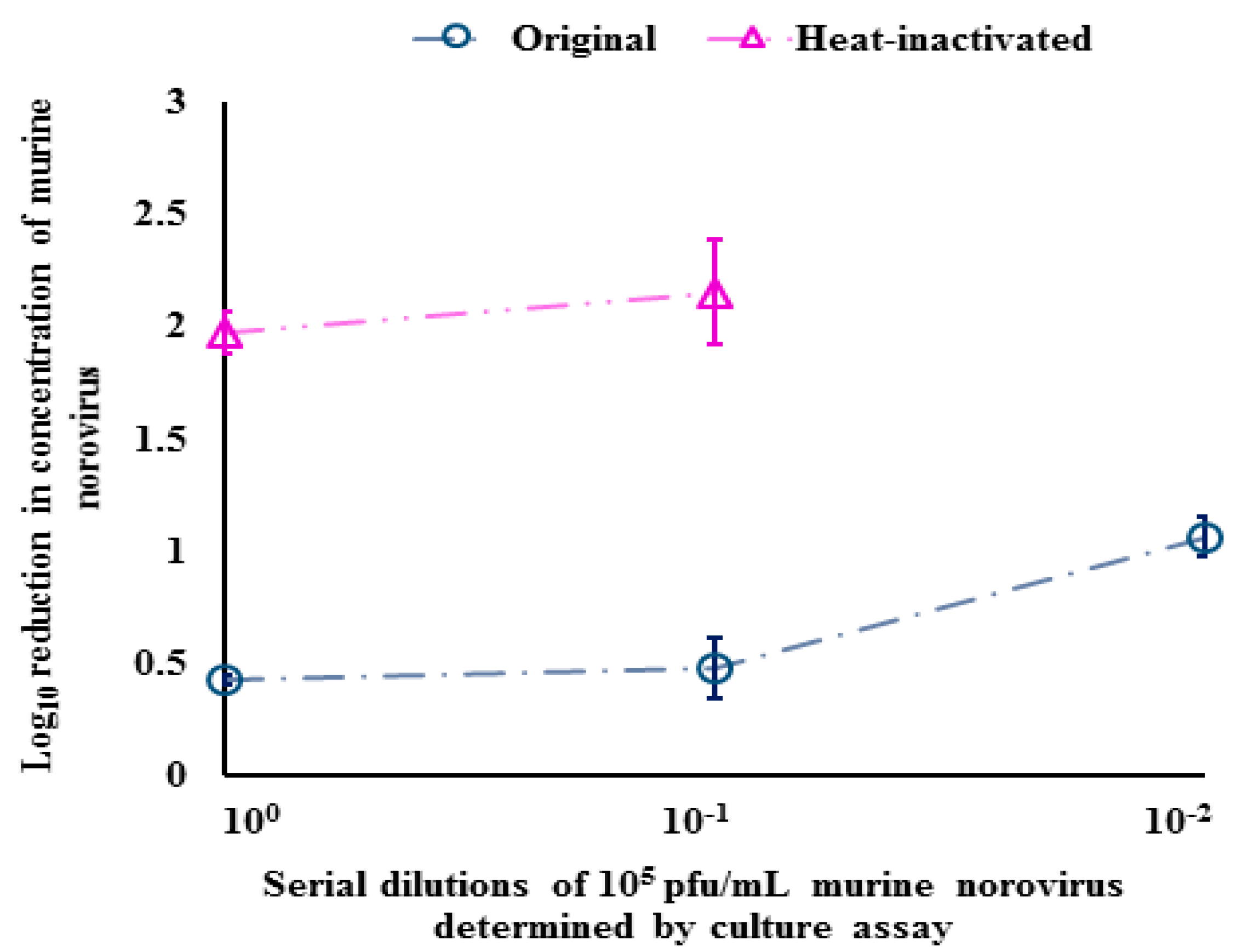
For heat-inactivated murine norovirus with no PEMAXTM treatment, the last dilution (10−4) was no longer detectable, with mean qPCR CT values of the positive samples ranging from 28.1 ± 0.1 (100 dilution) to 39.5 ± 0.2 (10−3 dilution). For these samples, the mean qPCR CT values obtained after PEMAXTM treatment were significantly (p < 0.05) higher (34.7 ± 0.2 (100 dilution) to 40.4 ± 0.4 (10−1 dilution), respectively, than those which were not treated with PEMAXTM (Figure 2). The PEMAXTM treatment resulted in a significant reduction (1.98 to 2.14 log10) of murine norovirus in heat-inactivated samples for the detected dilutions compared to the original (non-heat inactivated) samples (0.43 to 1.06 log10 reduction) (Figure 3).
3.3. Evaluation of PEMAXTM-RT-qPCR for the Selective Detection of Infectious Murine Norovirus
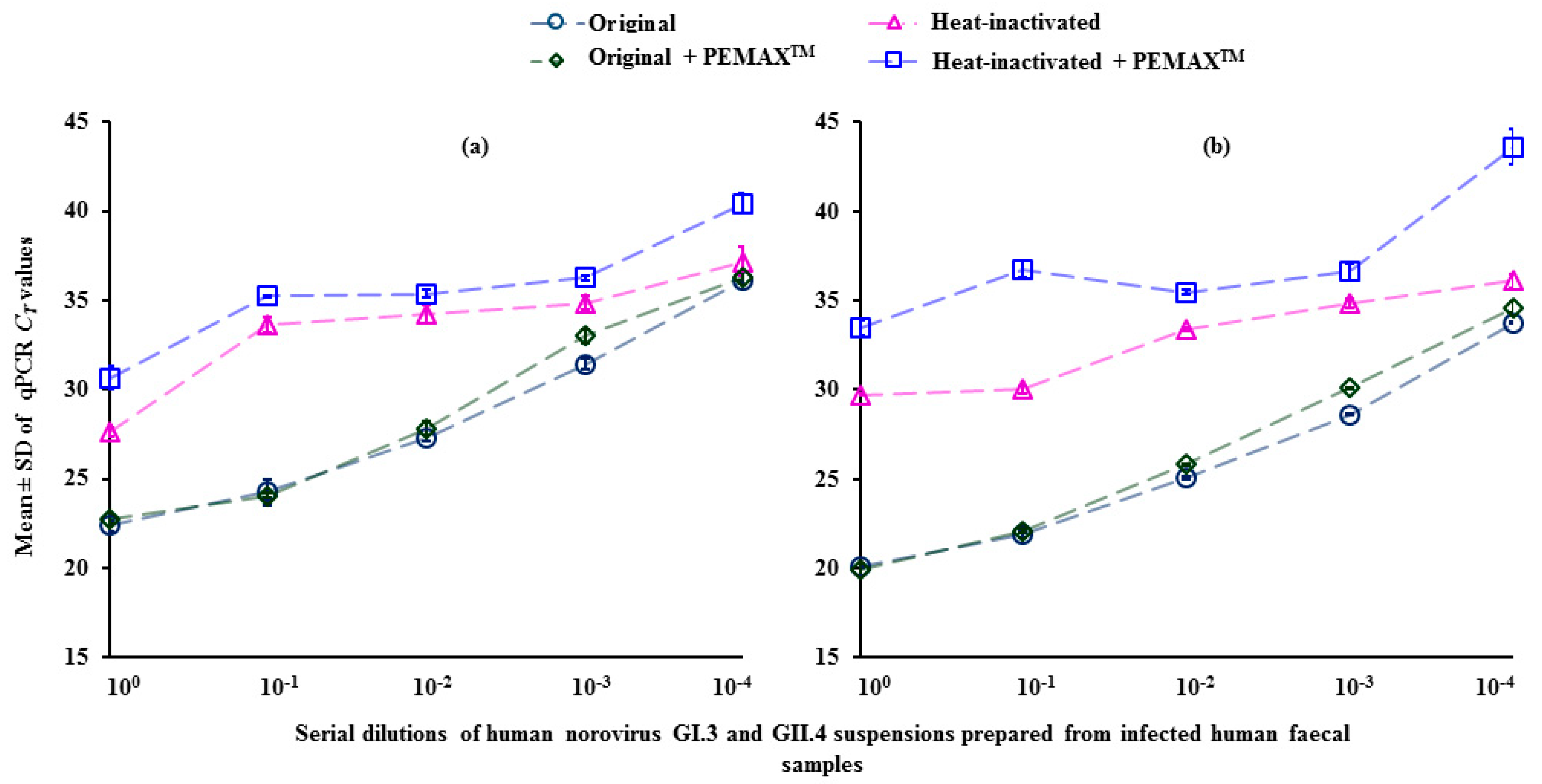
For original (non-heat-inactivated) norovirus GI.3, PEMAXTM treatment did not produce a significant difference in qPCR CT values (Figure 4a). However, there was a significant (p < 0.05) difference in qPCR CT values obtained for the heat-inactivated samples in all dilutions (100 to 10−4) after PEMAXTM treatment. The difference in the mean qPCR CT value was higher in the 10−4 (−3.3 ± 1.2) and 100 (−3.0 ± 1.0) dilutions. Similar results were observed for non-heat-inactivated norovirus GII.4 samples (Figure 4b). However, the difference in the mean qPCR CT value for heat-inactivated norovirus GII.4 samples with and without PEMAXTM treatment were greater than for GI.3. The difference in the mean qPCR CT value for heat-inactivated norovirus GII.4 samples with and without PEMAXTM treatment ranged from −7.5 ± 1.2 (10−4 dilution) to −1.8 ± 0.6 (10−3 dilution) (Figure 4b).
The PEMAXTM treatment resulted in a 0.34 to 0.98 log10 reduction of detectable norovirus GI.3 in heat-inactivated samples (Table 1). However, a significantly (p < 0.05) higher reduction (0.53 to 2.26 log10) was achieved for heat-inactivated norovirus GII.4 with the PEMAXTM treatment. The log10 reduction was greater when lower virus concentrations (i.e., 10−4 dilution) were used for both norovirus genotypes.
3.4. Application of PEMAXTM-RT-qPCR for Detection of Infectious Norovirus from Environmental Samples
A total of 10 samples [six influent wastewater (samples I1–I6), two effluent wastewater (samples E1 and E2) and two seawater (samples S1and S2)] were tested for norovirus GI and GII. With the exception of one influent wastewater sample (I4), that was negative for norovirus GI, all samples were positive for both norovirus GI and GII using RT-qPCR. For norovirus GI, PEMAXTM treatment resulted in its non-detection using RT-qPCR for both seawater samples (S1 and S2) compared to the PEMAXTM untreated samples (Table 2). The PEMAXTM treatment resulted in significantly higher qPCR CT values for both effluent (E1 and E2) and 60% (3/5) of the influent samples. Similar results were observed for norovirus GII. The PEMAXTM treatment resulted in complete elimination of qPCR amplification for one of the seawater samples (S2) and resulted in a significant increase in mean qPCR CT value for the other seawater sample (S1), both effluent samples and 50% (3/6) influent samples.
The analysis showed that although both seawater samples (S1 and S2) did not contain infectious norovirus GI, it did indicate that 44% of the norovirus GII detected in one seawater sample (S1) was infectious (Table 3). For the effluent samples E1 and E2, infectious norovirus GI (59% and 32%, respectively) and GII (24% and 52%, respectively) were detected. The analysis also indicated the presence of non-infectious norovirus (19 to 79% GI and 11 to 63% GII) in influent samples.
4. Discussion
Municipal wastewater potentially contains high concentrations of enteric viruses, and physical (sedimentation, activated sludge and trickling filters, irreversible adsorption) and chemical (disinfectant) treatment processes can be inefficient for their inactivation and/or removal [22,23]. Irrespective of wastewater treatment type, enteric viruses including noroviruses are likely to be present at concentrations up to 5 log10 genome copies in non-disinfected wastewater effluents. When virus-contaminated effluent is released into receiving environmental waters there are associated human health risks through environmental transmission [19,24]. Demonstration of virus infectivity in these waters is required to assess risk. To address the issue of RT-qPCR detecting non-infectious viruses, a norovirus PEMAXTM-RT-qPCR method was developed and evaluated for its applicability in the detection of infectious viruses from wastewater and seawater.
The photoactivatable dyes PMA and EMA have been used individually for the selective detection of infectious murine norovirus from heat-inactivated samples [8,13,25,26]. In those studies, PMA was less effective (0.14 to 1.38 log10 reduction) in discriminating between non-infectious and infectious murine norovirus [13,25] than shown by our results using PEMAXTM (~2.0 log10 reduction). This difference may be explained by the use of a 90 °C virus inactivation temperature in our study, much higher than the 65 °C and 72 °C temperatures used elsewhere [13,25]. As Millard and colleagues reported that complete inactivation of another enteric virus, hepatitis A virus, was only achieved when heated to 90 °C and maintained for 90 s [27], therefore, 90 °C for 3 min was chosen to ensure that complete capsid damage was achieved, with virus capsid would be likely permeable to the PEMAXTM dye [26].
Further studies should compare the effectiveness of PEMAXTM with other approaches, including methods that utilize an RNase pre-treatment to remove free viral RNA. In addition, different inactivation mechanisms (UV treatment, chlorination, etc.) should be explored because the effectiveness of photoactivable dye-based methods can provide different infectivity profiles [28].
Our results suggest that the effect of PEMAXTM was highest in samples containing a lower virus concentration. This finding is consistent with previous studies [25,29]. This could be that aggregation of inactivated virus particles would prevent entry of PEMAXTM through the damaged capsids [30]. In addition, more effective heat-inactivation may have been achieved in samples containing lower virus concentrations. Our study also indicated that complete discrimination of non-infectious norovirus using photoactivable dyes is difficult to achieve, as reported elsewhere [9,10,29]. This could be that the differential ability of dye-based RT-qPCR methods depend on the extent of capsid damage from heat-treatment. It has been reported that the extent and type of damage to the capsid of heat inactivated viruses is temperature dependent [13,30,31].
PEMAXTM was less effective on norovirus GI.3 (up to 1 log10 reduction detected) than for GII.4 (up to 2 log10). This may be associated with the finding that the structure of virus-like particles of GI is more heat resistant than GII [32]. However, our results contradict with the findings of a previous study where higher (0.41 log10) reductions for norovirus GI compared to GII (0.23 log10) were reported [9]. The minimal effect of PEMAXTM on that study could be associated with the lack of appropriate incubation step (i.e., 30 min in the dark) as recommended by the manufacturer. Therefore, direct comparisons between these two studies may not be feasible. Further inter-laboratory validation using a similar methodology and other norovirus genotypes would be beneficial to evaluate the efficiency of PEMAXTM for the selective detection of infectious viruses including noroviruses from environmental samples.
Finally, the efficiency of PEMAXTM for the selective detection of infectious norovirus in influent and effluent wastewater, and seawater was evaluated. Our results showed that we could not detect any infectious norovirus GI in seawater samples, possibly because of the low concentration of norovirus GI present in those samples. Similar results were reported in a previous study where 98% hepatitis A viruses present in river water were deemed non-infectious [33]. However, our results did indicate the low level of infectious norovirus GII present in one of the seawater samples.
Despite the significant reduction in infectious norovirus (GI and GII) in effluent wastewater, the PEMAXTM-RT-qPCR method indicated the presence of infectious noroviruses. The detection of noroviruses by the PEMAXTM-RT-qPCR method in these samples may reflect that ineffective virus inactivation and/or that capsid damage may not be sufficient to allow entry of the PEMAXTM, both resulting in a minimal effect of this approach. Despite limitations, the PEMAXTM-RT-qPCR method may serve as a better approach than RT-qPCR alone for the detection of noroviruses from environmental samples and can be useful when associated public health risks are to be evaluated. However, improvements are needed, including comparison of the PEMAXTM-RT-qPCR results with a norovirus in vitro cultivation method when available.
5. Conclusions
Until a reliable culture method for the assessment of norovirus infectivity is widely available, we have to rely on RT-qPCR methods, preferably modified with photoactivable dyes including PEMAXTM, PMA and EMA. In this study, we developed and evaluated the efficiency of PEMAXTM using RT-qPCR for the selective detection of infectious norovirus from naturally contaminated environmental samples including effluent wastewater. The PEMAXTM-RT-qPCR provided better results on infectivity of norovirus than RT-qPCR alone; however, the method has limitations as it did not completely eliminate RT-qPCR amplification from heat-inactivated norovirus. Despite the limitations, this approach has the potential to provide information on viral infectivity to assess the potential human health risks associated with food and water. However, further improvements of the PEMAXTM-RT-qPCR method and the use of different norovirus genotypes inactivated with different inactivation strategies such as UV treatment and chlorination should be considered before adapting for the routine use.
Author Contributions
Conceptualization, P.G. and J.H.; Methodology, P.G. and J.H.; Data Analysis, P.G.; Writing Original Manuscript and Review, P.G.; Editing Manuscript and Review, J.H.
Funding
This research was funded by the Ministry of Business, Innovation, and Employment (MBIE) (Safe New Zealand Seafood. CAWX1317 Project 15330).
Acknowledgments
Ministry of Business, Innovation, and Employment (MBIE) are acknowledged for providing research funding. The authors thank David Harte and Lucia Rivas for reviewing the manuscript and providing feedback, and Dawn Croucher for assistance with sample preparation. The authors thank Christiane E. Wobus, Department of Pathology and Immunology, Washington University School of Medicine, St. Louis, MO, USA for kindly donating murine norovirus.
Conflicts of Interest
The authors declare that there is no conflict of interest.
Ethical Statement
This research does not contain clinical studies or patient data.
References
- Hewitt, J.; Bell, D.; Simmons, G.C.; Rivera-Aban, M.; Wolf, S.; Greening, G.E. Gastroenteritis outbreak caused by waterborne norovirus at a New Zealand ski resort. Appl. Environ. Microbiol. 2007, 73, 7853–7857. [Google Scholar] [CrossRef] [PubMed]
- Le Guyader, F.S.; Parnaudeau, S.; Schaeffer, J.; Bosch, A.; Loisy, F.; Pommepuy, M.; Atmar, R.L. Detection and quantification of noroviruses in shellfish. Appl. Environ. Microbiol. 2009, 75, 618–624. [Google Scholar] [CrossRef] [PubMed]
- Cangelosi, A.G. , Meschke, S.J. Dead or alive: Molecular assessment of microbial viability. Appl. Environ. Microbiol. 2014, 80, 5881–5891. [Google Scholar] [CrossRef] [PubMed]
- Murakami, K.; Kurihara, C.; Oka, T.; Shimoike, T.; Fujii, Y.; Takai-Todaka, R.; Park, Y.; Wakita, T.; Matsuda, T.; Hokari, R.; et al. Norovirus binding to intestinal epithelial cells is independent of histo-blood group antigens. PLoS ONE 2013, 8, e66534. [Google Scholar] [CrossRef] [PubMed]
- Huang, P.; Farkas, T.; Zhong, W.; Tan, M.; Thornton, S.; Morrow, A.L.; Jiang, X. Norovirus and histo-blood group antigens: Demonstration of a wide spectrum of strain specificities and classification of two major binding groups among multiple binding patterns. J. Virol. 2005, 79, 6714–6722. [Google Scholar] [CrossRef] [PubMed]
- Ettayebi, K.; Crawford, S.E.; Murakami, K.; Broughman, J.R.; Karandikar, U.; Tenge, V.R.; Neill, F.H.; Blutt, S.E.; Zeng, X.; Qu, L.; et al. Replication of human noroviruses in stem cell-derived human enteroids. Science 2016, 353, 1387–1393. [Google Scholar] [CrossRef] [PubMed] [Green Version]
- Jones, M.K.; Watanabe, M.; Zhu, S.; Graves, C.L.; Keyes, L.R.; Grau, K.R.; Gonzalez-Hernandez, M.B.; Iovine, N.M.; Wobus, C.E.; Vinje, J.; et al. Enteric bacteria promote human and mouse norovirus infection of B cells. Science 2014, 346, 755–759. [Google Scholar] [CrossRef] [PubMed] [Green Version]
- Karim, M.R.; Fout, G.S.; Johnson, C.H.; White, K.M.; Parshionikar, S.U. Propidium monoazide reverse transcriptase PCR and RT-qPCR for detecting infectious enterovirus and norovirus. J. Virol. Methods. 2015, 219, 51–61. [Google Scholar] [CrossRef] [PubMed] [Green Version]
- Randazzo, W.; Lopez-Galvez, F.; Allende, A.; Aznar, R.; Sanchez, G. Evaluation of viability PCR performance for assessing norovirus infectivity in fresh-cut vegetables and irrigation water. Int. J. Food Microbiol. 2016, 229, 1–6. [Google Scholar] [CrossRef] [PubMed]
- Randazzo, W.; Khezri, M.; Ollivier, J.; Le Guyader, F.C.; Rodríguez-Díaz, J.; Aznar, R.; Sánchez, G. Optimization of PMAxx pretreatment to distinguish between human norovirus with intact and altered capsids in shellfish and sewage samples. Int. J. Food Microbiol. 2018, 266, 1–7. [Google Scholar] [CrossRef] [PubMed]
- Prevost, B.; Goulet, M.; Lucas, F.S.; Joyeux, M.; Moulin, L.; Wurtzer, S. Viral persistence in surface and drinking water: Suitability of PCR pre-treatment with intercalating dyes. Water Res. 2016, 91, 68–76. [Google Scholar] [CrossRef] [PubMed]
- Li, X.; Huang, R.; Chen, H. Evaluation of assays to quantify infectious human norovirus for heat and high-pressure inactivation studies using Tulane virus. Food Environ. Virol. 2017, 9, 314–325. [Google Scholar] [CrossRef] [PubMed]
- Leifels, M.; Jurzik, L.; Wilhelm, M.; Hamza, I.A. Use of ethidium monoazide and propidium monoazide to determine viral infectivity upon inactivation by heat, UV-exposure and chlorine. Int. J. Hyg. Environ. Health 2015, 218, 686–693. [Google Scholar] [CrossRef] [PubMed]
- Nocker, A.; Sossa, K.E.; Camper, A.K. Molecular monitoring of disinfection efficacy using propidium monoazide in combination with quantitative PCR. J. Microbiol. Methods 2007, 70, 252–260. [Google Scholar] [CrossRef] [PubMed]
- Nocker, A.; Cheung, C.Y.; Camper, A.K. Comparison of propidium monoazide with ethidium monoazide for differentiation of live vs. dead bacteria by selective removal of DNA from dead cells. J. Microbiol. Methods 2006, 67, 310–320. [Google Scholar] [CrossRef] [PubMed]
- Codony, F.; Agusti, G.; Allue-Guardia, A. Cell membrane integrity and distinguishing between active and inactive cells as a means of improving viability PCR. Mol. Cell Probes 2015, 29, 190–192. [Google Scholar] [CrossRef] [PubMed]
- Wobus, C.E.; Karst, S.M.; Thackray, L.B.; Chang, K.O.; Sosnovtsev, S.V.; Belliot, G.; Krug, A.; Mackenzie, J.M.; Green, K.Y.; Virgin, H.W. Replication of norovirus in cell culture reveals a tropism for dendritic cells and macrophages. PLoS Biol. 2004, 2, e432. [Google Scholar] [CrossRef] [PubMed] [Green Version]
- Hewitt, J.; Rivera-Aban, M.; Greening, G.E. Evaluation of murine norovirus as a surrogate for human norovirus and hepatitis A virus in heat inactivation studies. J. Appl. Microbiol. 2009, 107, 65–71. [Google Scholar] [CrossRef] [PubMed] [Green Version]
- Hewitt, J.; Leonard, M.; Greening, G.E.; Lewis, G.D. Influence of wastewater treatment process and the population size on human virus profiles in wastewater. Water Res. 2011, 45, 6267–6276. [Google Scholar] [CrossRef] [PubMed]
- Gyawali, P.; Sidhu, J.P.S.; Ahmed, W.; Jagals, P.; Toze, S. Rapid concentration and sensitive detection of hookworm ova from wastewater matrices using a real-time PCR method. Exp. Parasitol. 2015, 159, 5–12. [Google Scholar] [CrossRef] [PubMed] [Green Version]
- Kageyama, T.; Kojima, S.; Shinohara, M.; Uchida, K.; Fukushi, S.; Hoshino, F.B.; Takeda, N.; Katayama, K. Broadly reactive and highly sensitive assay for Norwalk-like viruses based on real-time quantitative reverse transcription-PCR. J. Clin. Microbiol. 2003, 41, 1548–1557. [Google Scholar] [CrossRef] [PubMed]
- Templeton, M.R.; Andrews, R.C.; Hofmann, R. Particle-associated viruses in water: Impacts on disinfection processes. Crit. Rev. Environ. Sci. Technol. 2008, 38, 137–164. [Google Scholar] [CrossRef]
- Charles, K.J.; Souter, F.C.; Baker, D.L.; Davies, C.M.; Schijven, J.F.; Roser, D.J.; Deere, D.A.; Priscott, P.K.; Ashbolt, N.J. Fate and transport of viruses during sewage treatment in a mound system. Water Res. 2008, 42, 3047–3056. [Google Scholar] [CrossRef] [PubMed] [Green Version]
- Hassard, F.; Sharp, J.H.; Taft, H.; LeVay, L.; Harris, J.P.; McDonald, J.E.; Tuson, K.; Wilson, J.; Jones, D.L.; Malham, S.K. Critical review on the public health impact of norovirus contamination in shellfish and the environment: A UK perspective. Food Environ. Virol. 2017, 9, 123–141. [Google Scholar] [CrossRef] [PubMed]
- Kim, S.Y.; Ko, G. Using propidium monoazide to distinguish between viable and nonviable bacteria, MS2 and murine norovirus. Lett. Appl. Microbiol. 2012, 55, 182–188. [Google Scholar] [CrossRef] [PubMed] [Green Version]
- Lee, M.; Seo, D.J.; Seo, J.; Oh, H.; Jeon, S.B.; Ha, S.D.; Myoung, J.; Choi, I.S.; Choi, C. Detection of viable murine norovirus using the plaque assay and propidium-monoazide-combined real-time reverse transcription-polymerase chain reaction. J. Virol. Methods 2015, 221, 57–61. [Google Scholar] [CrossRef] [PubMed]
- Millard, J.; Appleton, H.; Parry, J.V. Studies on heat inactivation of hepatitis A virus with special reference to shellfish. Epidemiol. Infect. 1987, 98, 397–414. [Google Scholar] [CrossRef] [PubMed]
- Li, X.; Chen, H. Evaluation of the porcine gastric mucin binding assay for high-pressure-inactivation studies using murine norovirus and tulane virus. Appl. Environ. Microbiol. 2015, 81, 515–521. [Google Scholar] [CrossRef] [PubMed]
- Parshionikar, S.; Laseke, I.; Fout, G.S. Use of propidium monoazide in reverse transcriptase PCR to distinguish between infectious and noninfectious enteric viruses in water samples. Appl. Environ. Microbiol. 2010, 76, 4318–4326. [Google Scholar] [CrossRef] [PubMed]
- Baert, L.; Wobus, C.E.; Van Coillie, E.; Thackray, L.B.; Debevere, J.; Uyttendaele, M. Detection of murine norovirus 1 by using plaque assay, transfection assay, and real-time reverse transcription-PCR before and after heat exposure. Appl. Environ. Microbiol. 2008, 74, 543–546. [Google Scholar] [CrossRef] [PubMed]
- Moore, M.D.; Bobay, B.G.; Mertens, B.; Jaykus, L.A. Human norovirus aptamer exhibits high degree of target conformation dependent binding similar to that of receptors and discriminates particle functionality. mSphere 2016, 1, e00298-16. [Google Scholar] [CrossRef] [PubMed]
- Samandoulgou, I.; Hammami, R.; Rayas, R.M.; Fliss, I.; Jean, J. Stability of secondary and tertiary structures of virus like particles representing noroviruses: Effects of pH, ionic strength, and temperature and implications for adhesion to surfaces. Appl. Environ. Microbiol. 2015, 81, 7680–7686. [Google Scholar] [CrossRef] [PubMed]
- Fuster, N.; Pinto, R.M.; Fuentes, C.; Beguiristain, N.; Bosch, A.; Guix, S. Propidium monoazide RTqPCR assays for the assessment of hepatitis A inactivation and for a better estimation of the health risk of contaminated waters. Water Res. 2016, 101, 226–232. [Google Scholar] [CrossRef] [PubMed] [Green Version]
Figure 1.
Differences in qPCR CT values (mean and standard deviation) for original (non-heat-inactivated) and heat-inactivated murine norovirus with and without PEMAXTM treatment.
Figure 1.
Differences in qPCR CT values (mean and standard deviation) for original (non-heat-inactivated) and heat-inactivated murine norovirus with and without PEMAXTM treatment.

Figure 2.
qPCR CT values (mean and standard deviation) of serially-diluted original (non-heat-inactivated) and heat-inactivated murine norovirus with and without PEMAXTM treatment.
Figure 2.
qPCR CT values (mean and standard deviation) of serially-diluted original (non-heat-inactivated) and heat-inactivated murine norovirus with and without PEMAXTM treatment.

Figure 3.
Log10 reduction (mean and standard deviation) of original (non-heat-inactivated) and heat-inactivated murine norovirus after PEMAXTM treatment.
Figure 3.
Log10 reduction (mean and standard deviation) of original (non-heat-inactivated) and heat-inactivated murine norovirus after PEMAXTM treatment.

Figure 4.
qPCR CT values (mean and standard deviation) of serially-diluted original (non-heat-inactivated) and heat-inactivated (a) norovirus GI.3 and (b) norovirus GII.4 with and without PEMAXTM treatment.
Figure 4.
qPCR CT values (mean and standard deviation) of serially-diluted original (non-heat-inactivated) and heat-inactivated (a) norovirus GI.3 and (b) norovirus GII.4 with and without PEMAXTM treatment.


Table 1.
Relative log10 reduction (mean and standard deviation) of norovirus GI.3 and GII.4 concentrations in original (non-heat-inactivated) and heat-inactivated suspensions after PEMAXTM treatment.
Table 1.
Relative log10 reduction (mean and standard deviation) of norovirus GI.3 and GII.4 concentrations in original (non-heat-inactivated) and heat-inactivated suspensions after PEMAXTM treatment.
| Dilutions | Relative log10 Reduction in Norovirus Concentration | |||
|---|---|---|---|---|
| GI.3 | GII.4 | |||
| Original | Heat-Inactivated | Original | Heat-Inactivated | |
| 100 | 0.11 ± 0.02 | 0.91 ± 0.16 | 0.05 ± 0.08 | 1.12 ± 0.13 |
| 10−1 | 0.05 ± 0.25 | 0.47 ± 0.12 | 0.06 ± 0.01 | 2.03 ± 0.08 |
| 10−2 | 0.17 ± 0.11 | 0.34 ± 0.21 | 0.13 ± 0.03 | 0.63 ± 0.07 |
| 10−3 | 0.17 ± 0.28 | 0.43 ± 0.13 | 0.15 ± 0.01 | 0.53 ± 0.03 |
| 10−4 | 0.05 ± 0.04 | 0.98 ± 0.35 | 0.25 ± 0.09 | 2.26 ± 0.02 |
Table 2.
Difference in qPCR CT values (mean and standard deviation) for norovirus GI and GII in wastewater and seawater samples with and without PEMAXTM treatment.
Table 2.
Difference in qPCR CT values (mean and standard deviation) for norovirus GI and GII in wastewater and seawater samples with and without PEMAXTM treatment.
| Sample Description | Sample ID | Norovirus GI | Norovirus GII | ||||
|---|---|---|---|---|---|---|---|
| qPCR CT Values (Mean ± SD) | ΔΔ CT | qPCR CT Values (Mean ± SD) | ΔΔ CT | ||||
| (−) PEMAXTM | (+) PEMAXTM | (−) PEMAXTM | (+) PEMAXTM | ||||
| Influent wastewater | I1 | 34.9 ± 0.24 | 36.6 ± 0.73 | −1.65 | 33.2 ± 0.61 | 33.8 ± 0.36 | −0.53 |
| I2 | 36.5 ± 0.60 | 38.6 ± 0.15 | −2.14 | 28.1 ± 0.04 | 29.5 ± 0.07 | −1.42 | |
| I3 | 34.9 ± 0.28 | 35.2 ± 0.23 | −0.30 | 27.2 ± 0.07 | 27.4 ± 0.12 | −0.17 | |
| I4 | ND | ND | NA | 40.6 ± 1.01 | 40.9 ± 0.67 | −0.22 | |
| I5 | 37.5 ± 0.10 | 39.8 ± 0.23 | −2.23 | 31.5 ± 0.07 | 32.6 ± 0.14 | −1.15 | |
| I6 | 30.4 ± 0.36 | 31.9 ± 0.10 | −1.42 | 31.4 ± 0.01 | 32.7 ± 0.23 | −1.29 | |
| Effluent wastewater | E1 | 39.6 ± 0.32 | 40.4 ± 0.10 | −0.76 | 37.2 ± 0.26 | 39.3 ± 0.98 | −2.05 |
| E2 | 33.1 ± 0.26 | 34.7 ± 0.06 | −1.63 | 28.3 ± 0.09 | 29.3 ± 0.12 | −0.94 | |
| Seawater | S1 | 40.8 ± 0.59 | ND | NA | 37.8 ± 0.36 | 39.0 ± 0.04 | −1.12 |
| S2 | 40.2 ± 1.05 | ND | NA | 37.4 ± 0.39 | ND | NA | |
ΔΔ CT = Difference in qPCR CT values. ND = not detected. NA = not applicable. Statistically significant (p < 0.05) results are shown in bold.
Table 3.
Percentage of infectious norovirus GI and GII in wastewater and seawater samples as determined by PEMAXTM-RT-qPCR.
Table 3.
Percentage of infectious norovirus GI and GII in wastewater and seawater samples as determined by PEMAXTM-RT-qPCR.
| Sample Description | Sample ID | Percentage of Infectious Norovirus Detected | |
|---|---|---|---|
| GI | GII | ||
| Influent wastewater | I1 | 32 | 59 |
| I2 | 23 | 37 | |
| I3 | 81 | 89 | |
| I4 | NA | 86 | |
| I5 | 21 | 45 | |
| I6 | 38 | 41 | |
| Effluent wastewater | E1 | 59 | 24 |
| E2 | 32 | 52 | |
| Seawater | S1 | 0 | 44 |
| S2 | 0 | 0 | |
NA = Not applicable (as norovirus GI not detected by RT-qPCR).
© 2018 by the authors. Licensee MDPI, Basel, Switzerland. This article is an open access article distributed under the terms and conditions of the Creative Commons Attribution (CC BY) license (http://creativecommons.org/licenses/by/4.0/).
Share and Cite
MDPI and ACS Style
Gyawali, P.; Hewitt, J. Detection of Infectious Noroviruses from Wastewater and Seawater Using PEMAXTM Treatment Combined with RT-qPCR. Water 2018, 10, 841. https://doi.org/10.3390/w10070841
AMA Style
Gyawali P, Hewitt J. Detection of Infectious Noroviruses from Wastewater and Seawater Using PEMAXTM Treatment Combined with RT-qPCR. Water. 2018; 10(7):841. https://doi.org/10.3390/w10070841
Chicago/Turabian StyleGyawali, Pradip, and Joanne Hewitt. 2018. "Detection of Infectious Noroviruses from Wastewater and Seawater Using PEMAXTM Treatment Combined with RT-qPCR" Water 10, no. 7: 841. https://doi.org/10.3390/w10070841
Note that from the first issue of 2016, this journal uses article numbers instead of page numbers. See further details here.